Download
1 / 50
510 likes | 836 Views
Cancer Stem Cells and Metastasis. Maria M. (Marj) Pe ña, PhD Dept. of Biological Sciences Center for Colon Cancer Research University of South Carolina. Normal stem cells.

E N D
Cancer Stem Cells and Metastasis Maria M. (Marj) Peña, PhD Dept. of Biological Sciences Center for Colon Cancer Research University of South Carolina
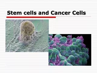
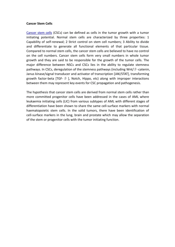
Normal stem cells Rare cells within organs with the ability to self-renew and give rise to all types of cells within the organ to drive organogenesis Cancer stem cells Rare cells within tumors with the ability to self-renew and give rise to the phenotypically diverse tumor cell population to drive tumorigenesis
Properties shared by normal stem cells and cancer stem cells • Assymetric Division: • Self renewal • Tissue-specific normal stem cells must self-renew throughout the lifetime of the animal to maintain specific organs • Cancer stem cells undergo self-renewal to maintain tumor growth • Differentiation into phenotypically diverse mature cell types • Give rise to a heterogeneous population of cells that compose the organ or the tumor but lack the ability for unlimited proliferation (hierarchical arrangement of cells) • Regulated by similar pathways • Pathways that regulate self-renewal in normal stem cells are dys-regulated in cancer stem cells
Development of Hematopoietic Stem Cells Liquid Organ Stem Cells Multipotent Progenitors Oligolineage Progenitors Mature Cells HSCs can be subdivided into long-term self-renewing HSCs, short-term self-renewing HSCs and multipotent progenitors (red arrows indicate self-renewal). They give rise to common lymphoid progenitors (CLPs; the precursors of all lymphoid cells) and common myeloid progenitors (CMPs; the precursors of all myeloid cells). Both CMPs/GMPs (granulocyte macrophage precursors) and CLPs can give rise to all known mouse dendritic cells. ErP, erythrocyte precursor; MEP, megakaryocyte erythrocyte precursor; MkP, megakaryocyte precursor; NK, natural killer. Differentiation Self Renewal Reya et al. 2001 Nature 414:105-111
Stem Cells Multipotent Progenitors Oligolineage Progenitors Mature Cells How do we identify Hematopoietic Stem Cells CD34- CD38+ CD20+ CD8+ CD8+ CD34+ CD38- CD4+ CD34- CD38- CD4+ CD36+ CD35+ Reya et al. 2001 Nature 414:105-111
Anti-A Antibody Anti-B Antibody
CD34 Expression CD38 Expression Self-renewal Assay Irradiated Mice FACS Cell Sorter Bone Marrow Cells Lethally irradiated Mice
Stem Cells Multipotent Progenitors Oligolineage Progenitors Mature Cells How do we identify Hematopoietic Stem Cells CD34- CD38+ CD20+ CD8+ CD8+ CD34+ CD38- CD34- CD38- CD4+ CD4+ CD36+ CD35+ Reya et al. 2001 Nature 414:105-111
The anatomy of the small intestinal epithelium Differentiation Self Renewal The epithelium is shaped into crypts and villi (left). The lineage scheme (right) depicts the stem cell, the transit-amplifying cells, and the two differentiated branches. The right branch constitutes the enterocyte lineage; the left is the secretory lineage. Relative positions along the crypt-villus axis correspond to the schematic graph of the crypt in the center. F. Radtke et al., Science 307, 1904 -1909 (2005)
Adult intestinal homeostasis Schematic representation and section of the crypt-villus unit in the mature small intestine. Proliferative cells reside in the crypts, while differentiated cells occupy the villus. Crypt progenitors migrate up (red arrow) the crypt-villus axis before shedding into the lumen. The process of epithelial renewal takes 3-6 d and is ensured by a small number of asymmetrically dividing stem cells at the bottom of the crypts. Wnt signaling in the adult intestine promotes proliferation of progenitor or transit-amplifying (TA) cells, as well as commitment toward secretory lineages. Wnt signaling may also drive terminal differentiation of certain secretory lineages. Although it is commonly believed that Wnt signaling may promote proliferation and/or differentiation of intestinal stem cells, there is no evidence that formally proves this (see arrows with question marks). In panel A, black arrowheads indicate Ki67 positive transit-amplifying cells, while white arrowheads indicate the Paneth cell compartment. Alex Gregorieff et al. Genes Dev. 2005; 19: 877-890
Pathways involved in self-renewal that are deregulated in cancer cells Wnt, Shh, and Notch pathways have been shown to contribute to the self-renewal of stem cells and/or progenitors in a variety of organs, including the haematopoietic and nervous systems. When dysregulated, these pathways can contribute to oncogenesis. Mutations of these pathways have been associated with a number of human tumours, including colon carcinoma and epidermal tumours (Wnt), medulloblastoma and basal cell carcinoma (Shh), and T-cell leukaemias (Notch).
Origin of the Theory of Cancer Stem Cells • Only a small subset of cancer cells is capable of • extensive proliferation • Liquid Tumors • In vitro colony forming assays: • - 1 in 10,000 to 1 in 100 mouse myeloma cells obtained from ascites away from normal hematopoietic cells were able to form colonies • In vivo transplantation assays: • - Only 1-4% of transplanted leukaemic cells could form spleen colonies • Solid Tumors • - A large number of cells are required to grow tumors in xenograft models • - 1 in 1,000 to 1 in 5,000 lung cancer, neuroblastoma cells, ovarian cancer cells, or breast cancer cells can form colonies in soft agar or in vivo
Two General Models for Cancer Heterogeneity 1. All cancer cells are potential cancer stem cells but have a low probability of proliferation in clonogenic assays 2. Only a small definable subset of cancer cells are cancer stem cells that have the ability to proliferate indefinitely.
Self renewal and differentiation are random. All cells have equal but low probability of extensive proliferation. Only cells with self renewal capacity can sustain tumor growth. Distinct classes of cells exist within a tumor. Only a small definable subset, the cancer stem cells can initiate tumor growth.
Therapeutic implications of Cancer Stem Cells • Most therapies fail to consider the difference in drug sensitivities of cancer stem cells • compared to their non-tumorigenic progeny. • Most therapies target rapidly proliferating non-tumorigenic cells and spare the • relatively quiescent cancer stem cells.
Thymidylate synthase Chu E. et al., Cancer Chemother Pharmacol (2003) 52 (Suppl 1) S80-S89
Thymidylate Synthase Inhibitors Raltitrexed 5-FU Longley, DB et al., Nature Reviews Cancer (2003) 3:330-338
CD34 Expression CD38 Expression Self-renewal Assay in NOD/SCID Mice (Non-obese diabetic/severe combined immunodeficiency) FACS Cell Sorter Cancer Cells ex: Leukaemia cells Sublethally irradiated NOD/SCID Mice
Hierarchies in normal and leukemic human hematopoietic cells Human hematopoietic cells are organized in a hierarchy that is sustained by a small population of self-renewing hematopoietic stem cells (HSCs). HSCs give rise to progressively more lineage-restricted, differentiated progenitors with reduced self-renewal capacity (LTC-ICs, long-term culture-initiating cells; CFU, colony-forming units), which in turn produce functionally mature blood cells. Disruption of pathways regulating self-renewal and differentiation through the acquisition of transforming mutations generates leukemic stem cells (LSCs) capable of sustaining growth of the leukemic clone in vivo. LSCs possess an altered differentiation program, as demonstrated by aberrant expression of some cell-surface markers (indicated in blue) and give rise to an aberrant developmental hierarchy that retains aspects of its normal counterpart. In vivo reconstitution assays using immune-deficient mouse recipients enable detection of HSCs and LSCs as SCID-repopulating cells (SRCs) and SCID leukemia-initiating cells (SL-ICs), respectively. Wang and Dick 2005 Trends in Cell Biology 15:494-501
The importance of self-renewal in leukemic initiation and progression. Self-renewal is a key property of both normal and leukemic stem cells. Fewer mutagenic changes are required to transform stem cells in which the self-renewal machinery is already active (a), as compared with committed progenitors in which self-renewal must be activated ectopically (b). In addition, self-renewing stem cells are long-lived; thus, there is an increased chance for genetic changes to accumulate in individual stem cells in comparison with more mature, short-lived progenitors. If a committed progenitor with limited life span acquires a genetic mutation that does not confer increased self-renewal (c), that cell will likely die or undergo terminal differentiation before enough mutations occur to propagate a full leukemogenic program.
Hematopoietic Cancer Stem Cells Acute myeloid leukemia (AML) –CD34+ CD38- Leukaemic Mouse Models: chronic myelomonocytic leukaemia (CMML) MRP8-BCL-2 acute myeloid leukaemia (AML) MRP8-BCL2Xlpr/lpr chronic myeloid leukaemia (CML)/BlastMRP8-PML-RARα acute promyelocytic leukaemia (APML)77 MRP8-BCRablXBCL-2
CD44 Expression CD24 Expression Self-renewal Assay in NOD/SCID Mice For solid tumors: surgical orthotopic implantation (SOI) FACS Cell Sorter Single Cell Suspension Solid Tumor Mince (small pieces) Surgical Implantation
Brain Tumor Stem Cells: CD133+ CD133 – neuronal stem cell marker Brain tumor stem cells were identified from human brain tumor samples by in vitro neurosphere assays normally used to isolate normal neural stem cells GFAP = glial fibrillary acidic protein Singh et. al 2003 Cancer Research 63: 5821-5828.
CD133+ CD133+ CD133- Brain tumor stem cells were identified by intracranial transplantation of CD133+ cells into adult NOD/SCID mouse forebrain. Singh et al. 2004 Nature 432: 396-401
Breast Cancer Stem Cells:CD44+ CD24low Lin- B38.1+ ESA+ CD44 and CD24 – adhesion molecules B38.1 – breast/ovarian cancer-specific marker ESA – epithelial specific antigen Al-Hajj, Muhammad et al. (2003) Proc. Natl. Acad. Sci. USA 100, 3983-3988
FUTURE DIRECTIONS • Need to characterize cancer stem cells at the single cell level • Understand the genetic and biochemical mechanisms that • control the self-renewal phenotype, assymetric subdivision, and the role of the stem cell niche in regulating the biological properties of both normal and cancer stem cells. • Characterize the response of cancer stem cells to • chemotherapeutic regimens • Develop therapeutic strategies to target cancer stem cells to prevent tumor recurrence.
Metastasis • Process by which a tumor cell leaves the primary tumor, travel to a distant site via the circulatory system and then establishes a secondary tumor. • Final and most devastating step of a malignancy • Leading cause of death in cancer patients • Before mets tumors may be cured by surgery
Metastasis is a multi-step processMetastatic cell = “Decathlon champion” Vascularization of primary tumor Tumor grows through the synthesis and secretion of pro-angiogenic factors by the tumor and surrounding stroma Invasion of the organ stroma through enhanced expression of enzymes (MMP9) Invasion of the lymphatic or vascular channels (may grow in these places) Tumors cells enter circulation Must survive turbulence of circulation and evade both immune and non-immune mechanisms
Metastatic Cancer Cells = Migratory Cancer Stem Cells • Cells arrest in the capillary beds of distant organs • Extravasation into distant organ • Survival and proliferation in target organ • Depends on multiple interactions (“cross-talk”) between tumor cell and organ microenvironment
Metastasis is not random • Seed and soil hypothesis • 1889: Stephen Paget analyzed autopsy records of 735 women with breast cancer • Metastasis to distant sites was not due to chance • Certain tumor cells (the “seed”) has an affinity for the milieu (the “soil”) of certain organs. Metastases resulted when the seed and soil were compatible • Metastatic dissemination occurs by purely mechanical factors that are the result of the anatomical structure of the vascular system • 1929: J. Ewing • Regional metastases can be attributed to anatomic and mechanical factors but distant organ metastases is specific • 1964: Sugarbaker • Lymphatic drainage to regional lymph nodes • Organ-specific metastases: breast, prostate, and lung cancer metastasize to the bone, while colorectal cancer metastasized to the liver and lymph nodes
Principles of the Seed and Soil Hypothesis • Tumors are biologically heterogeneous and contain subpopulations of cells with different angiogenic, invasive, and metastatic properties. • Metastases is a selective process for cells that succeed in invasion, embolization, survival in the circulation, arrest in a distal capillary bed, extravasation into the distant organ, and survival and proliferation in the distant organ. • The outcome of metastasis depends on multiple interactions (“cross-talk”) between the metastatic subpopulation in the primary tumor and the host organ microenvironment.
Tumors are biologically heterogeneous and contain subpopulations of cells with different angiogenic, invasive, and metastatic properties.
Organ Specific metastasis of Breast Cancer Cells MDA-MB-231 Breast Cancer Cell Line Isolate Single Clonal Populations (SCPs) Introduce Luciferase Bioluminescent Marker and GFP Fluorescence Marker Introduce into Nude Mice by intracardiac Injection Minn, A. J. et al. J. Clin. Invest. 2005;115:44-55
Noninvasive BLI to monitor the development of osteolytic metastases from the same mouse
Verification of macroscopic and microscopic metastases by fluorescence histology
SCPs exhibit different abilities to metastasize to bone
SCPs demonstrate different abilities to metastasize to the lung
Metastases is a selective process for cells that succeed in invasion, embolization, survival in the circulation, arrest in a distal capillary bed, extravastion into the distant organ, and survival and proliferation in the distant organ.
SCPs from MDA-MB-231 cells have a poor-prognosis gene expression signature Minn, A. J. et al. J. Clin. Invest. 2005;115:44-55
Genes that mediate metastasis to the Bone CXCR4 – bone homing chemokine receptor CTGF – connective tissue growth factor IL-11 – activator of osteoclast differentiation (mediators of bone resorption in bone metastases) MMP1 – matrix metalloproteinase/collagenase, promotes osteolysis by cleaving a specific peptide bond in the collagen of bone matrix OPN – osteopontin (consistently overexpressed in metastatic cells)
Genetic determinants for metastasis to the bone
The outcome of metastasis depends on multiple interactions (“cross-talk”) between the metastatic subpopulation in the primary tumor and the host organ microenvironment.
FUTURE DIRECTIONS • Understand the factors and mechanisms that lead to metastasis rather than study metastatic end points • What steps of metastasis provides good therapeutic targets? • Are the early steps clinically detectable and is the process a good biological target? • Understand the “cross-talk’ between metastatic cells and target organs that establish metastases • What are the “messages” • What are the “messengers” • Target the soil to prevent the growth of the seed • Develop therapies to alleviate metastases while minimizing therapies that will subject the patient to unnecessary toxicities
Wnt Signaling Pathway Fodde, R et al., Nat Rev Cancer (2001) 1:57-67