Download
1 / 40
450 likes | 923 Views
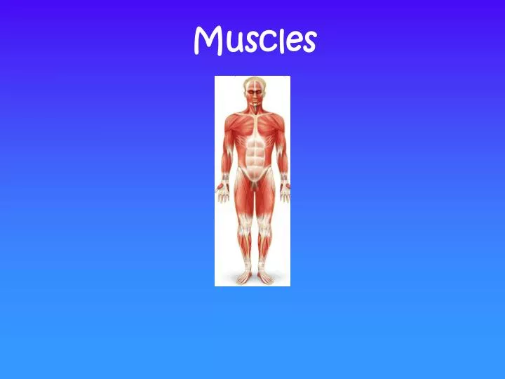
Muscles. What you Need to Know. Look briefly at the Structure of: 1) Skeletal, 2) Smooth & 3) Cardiac Muscle Naming, Identification, Functions You are only responsible for the identification of ALL muscles which are on your Lab Packet In general:

E N D
What you Need to Know • Look briefly at the Structure of: 1) Skeletal, 2) Smooth & 3) Cardiac Muscle • Naming, Identification, Functions You are only responsible for the identification of ALL muscles which are on your Lab Packet In general: Generates force to move your bones Propel body fluids & ingested food Generate and distribute heat
Specialized for contraction movement of skeleton or internal organs c) smooth a) skeletal b) cardiac Muscle
-generally attaches to bone causes movement of skeleton Characteristics: -long & unbranched, tightly packed, parallel cells Skeletal Muscle -multinucleated -striated (striped) -voluntary (conscious control)
-muscle of the heart Characteristics: -1 nucleus per cell Cardiac Muscle -branched cells with mild striations -involuntary (not consciously controlled)
-movement within internal organs other than the heart (e.g. digestive tract, bladder, arteries, etc.) Characteristics: Smooth Muscle -tapered cells -single nucleus/cell -no striations -involuntary
Dense Regular Connective Tissue -forms tendons (attach muscle to bone)and ligaments (attach bone to bone) -fibroblasts in collagen matrix Collagen fibers Fibroblasts
Facial Muscles p. 84 Frontalis: raises eyebrows; wrinkles forehead Temporalis: elevates mandible Orbicularis oculi: closes eye Buccinator: compresses cheek Orbicularis oris: closes and purses lips Masseter: elevates mandible (closes jaw)
Facial Muscles Frontalis: raises eyebrows; wrinkles forehead Temporalis: elevates mandible Orbicularis oculi: closes eye Orbicularis oris: closes and purses lips Buccinator: compresses cheek Masseter: elevates mandible (closes jaw) No equivalent picture in lab manual
Sternocleidomastoid -If both contracted flexion of head when both (nod down) -If one contracted rotates head to opposite shoulder
Muscles Many muscles form antagonistic muscle pairs Agonist: muscle that’s causing movement Antagonist: muscle that opposes the agonist An example… During arm flexion: Agonist: Biceps brachii Antagonist: Triceps brachii During arm extension: Agonist: Triceps brachii Antagonist: Biceps brachii
Muscles that Move Shoulder Trapezius: elevates (i.e. raises shoulders) & retracts scapula (i.e. pulls shoulders back) Elevates scapula Retracts scapula
Muscles that Move Shoulder No pic in lab manual Pectoralis minor: draws scapula forward & downward (i.e. pulls shoulders down and forward) Draws scapula forward Draws scapula downward
Muscles that Move Shoulder p. 82, 85 Serratus anterior: draws scapula forward (i.e. pulls shoulder forward) Serratus anterior Draws scapula forward
Muscles that Move the Upper Arm Pectoralis major: flexes (moves arm up and forward) & adductsupper arm (moves arm toward the body) Flexes upper arm Adducts upper arm
Muscles that Move the Upper Arm Latissimus dorsi: extends (i.e. moves arm down to the rear) & adducts arm (moves arm toward body) Extends upper arm Adducts upper arm
Muscles that Move Upper Arm Deltoid Deltoid: abducts arm (raises arm up away from the side) Abducts arm
Moving the Forearm Biceps brachii: flexes forearm Brachialis: flexes forearm Flexion of forearm Biceps brachii Brachialis Biceps brachii Brachialis
Muscles that Move the Forearm p. 83, 85 Triceps brachii: extends the forearm extension
Flexors & Extensors of Hand Hand flexor group: flex the hand p. 82. 84, 85 Hand extensor group: extend the hand p. 83. 85
Muscles of Abdominal Wall –Movement of Spine p. 82 Rectus abdominus: flexes vertebral column
Muscles of Abdominal –Moving the spine Lateral flexion External oblique: compresses abdomen; rotates trunk; lateral flexion Trunk Rotation Internal oblique: compresses abdomen; trunk rotation; lateral flexion No pic Compressing abdomen Transversus abdominis: compresses abdomen No pic
Muscles of Chest Wall –Movement of Ribs Internal intercostals: depresses rib cage during forced expiration External intercostals: elevates rib cage during inspiration No pics Diaphragm: prime mover during inspiration; increases vertical dimension of thorax
Muscles of Chest Wall –Movement of Ribs Internal intercostal External intercostal
Muscles that Move the Thigh Gluteus maximus: extends thigh p. 83, 86
Muscles that Move the Thigh iliopsoas: flexion of thigh Flexion of thigh
Muscles that Move the Thigh Flexion of thigh Tensor fascia latae: flexes & abducts thigh Abducts of thigh
Muscles that Move the Thigh Adductor group: adducts and flexes thigh Adducts of thigh Flexion of thigh
Muscles that Move the Thigh sartorius: flexes & laterally rotates thigh; helps you sit cross-legged Lateral rotation & flexion of thigh
Muscles that Move the Thigh Knee extension Quadriceps femoris group: extends knee Rectus femoris: extends knees and flexes thigh Flexion of thigh
Muscles that Move the Lower Leg Extension of thigh Hamstring group: extends thigh & flexes knee Knee flexion
Muscles that Move the Foot Tibialis anterior: dorisflexes foot Gastrocnemius: plantar flexes foot, flexes knee Soleus: plantar flexes foot
Today’s Activities Muscles: Tissue & Histology Examine the following slides using the microscope: 1) Skeletal muscle 3) Cardiac muscle 4) Dense regular connective tissue 2) Smooth muscle Muscles: Gross Anatomy Use the models and diagrams in you textbook & lab manual to study locations & actions of the muscles listed on pp. 76-79 Diagrams in textbook = right side; most models = left side Don’t worry about extra muscles described in textbook Don’t worry about extra actions described in textbook Answer questions, fill out diagrams on pp. 80-86 If you get done early, you can go back to the bones, etc. DO NOT leave until you have been excused, or you will be marked absent!!!
Medial View Radial view Medial View-Deep flexors Lateral View Lateral View-Deep extensors Lateral View-Deep shoulder extensors
Anterior View Posterior View Lateral View Medial View Plantar surface