Download
1 / 24
250 likes | 347 Views
Microcontrol of neuronal outgrowth. HELEN M. BUETTNER ( Nanofabrication and biosystems ) Student : Jiun-Shiung Feng Advisor : M.S. Ju C.C. K. Lin. The Neuron. Growth cone. Neurite outgrowth is guided by the growth cone .

E N D
Microcontrol of neuronal outgrowth HELEN M. BUETTNER ( Nanofabrication and biosystems ) Student : Jiun-Shiung Feng Advisor : M.S. Ju C.C. K. Lin
Growth cone • Neurite outgrowth is guided by the growth cone. • The growth cone consists of thin filopodia and lamellipodial veils.
Filopodial contact with remote cues can initiate rapid neurite advance to the point of contact. (O’Connor et al. 1990;Myers and Bastiani, 1993) • Proper placement of such cues in series reproduces stereotyped pathways. (Caudy and Bentley, 1986) • A model for growth cone behavior. (Buettner et al., 1994)
Mechanisms of growth cone advance (O’Connor et al. 1990) • Lamellipodial advance • The forward movement of the growth cone occur primarily through the net forward flow of lamellipodial veils. • Filopodial dilation (Guideposting) • filopodial can detect a region of favorable substrate (guidepost) and cause the growth cone to move toward it.
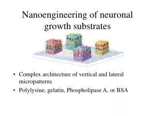
Microstructural paradigm • Use pairs of substrates • Permissive-Nonpermissive substrate • Laminin-glass (Clark et al., 1993) • Laminin-albumin (Hammarback and Letourneau,1986) • Laminin-collagen (Gundersen, 1987) • Laminin:a kind of glycoprotein, a major component of basement membranes
Quantitative framework (1/2) • Two key periods of advance • Movement across a homogeneous region according to the random walk characteristics of lamellipodial advance. • Filopodial dilation may across the nonpermissive region once filopodial contact has been made.
Quantitative framework (2/2) • is the probability that a growth cone at the border will detect and respond to a neighboring permissive region • is the number of contacting filopodia • is the critical threshold
Experimental Methods • Instrument (Buettner, 1994) • Microscope (100x objective) • Video camera • Videocassette(1 frame/sec ) • Digitized as 8-bit computer image (256 gray levels) • Outline is stored as binary image
Experimental Methods • Lamellipodial advance is characterized by tracking the geometrical center of lamellipodial region. y x
Experimental Methods • Filopodial dynamics • Filopodial initiation Initiation of filopodia appearing on a growth cone during the observation sequence (Poisson event) • Filopodial extension and retraction Measure by plotting the trajectories of individual filopodium tips on given growth cone (filopodial length)
Experimental Methods • Filopodial length • The straight-line distance between the position of the filopodium tip at any time and its initial position
Experimental Results • Lamellipodial advance The random term can be rewritten as N(0,1) represents a random variable taken from a normal distribution with a mean of zero and a variance of 2t. (berg, 1983)
Experimental Results • Filopodial dynamics Parameters of filopodial dynamics • Rate of initiation, • Rates of extension and retraction, re and rr • Maximun length, Lmax
Quantitative growth cone response to micropatterned environment • Time to reach a border • Mean time for a neurite to track a distance L
Summary and Conclusions • Model: • Probabilistic events between growth cone filopodia and microstructure feature. • The random walk advance of growth cone. • Application: • repair of nerve injuries. • construction of next-generation bioartificial organs.