Download
1 / 24
240 likes | 254 Views
MICROSCOPY AND HISTOCHEMISTRY. HISTOLOGICAL TECHNIQUE A. Histology involves the preparation of tissues for examination with a microscope. 1. Basic methods of histological preparation of tissues. a. Fix tissue (e.g. 4 % paraformaldehyde + buffer)

E N D
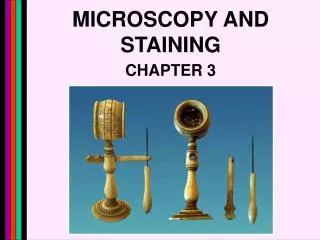
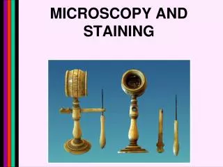
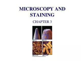
MICROSCOPY AND HISTOCHEMISTRY
HISTOLOGICAL TECHNIQUE A. Histology involves the preparation of tissues for examination with a microscope. 1.Basic methods of histological preparation of tissues. a. Fix tissue (e.g. 4 % paraformaldehyde + buffer) b. Dehydrate tissue (alcohol series followed by toluene) c. Embed tissue in “hard” medium (e.g. wax) d. Section embedded tissue on a microtome e. Mount sections on a supportive structure (e.g. slide) that can be placed on a microscope stage f. Usually remove the embedding medium g. Stain tissue (e.g. hematoxylin-eosin) h. Examine tissue with microscope.
2. Why fix tissue? a. Preserve structure. Essentially to make the structural components of the tissue more durable so that the tissue can be manipulated in various ways. • Fixed material is dead. You want to preserve the structure (chemical and morphological) of the living material so that it appears the same as it was in life. • It will never be exactly the same. Important to choose fixative that does the best job. Fixative used will depend on type of tissue to be fixed. 3. Why dehydrate the fixed tissue a. Most fixatives are water soluble, most embedding media are non-polar and are not miscible with water. So, you have to move the tissue from a polar (water-based) medium to a non-polar medium (e.g. toluene) that is miscible with the embedding medium.
4. Why embed? a. Tissue will be sectioned. Needs to be durable enough to withstand the sectioning process. Also, want components of tissue to remain in their natural positions. Don't want them to be moved to new positions. b. Embedding in wax or plastic immobilizes structural components of tissue. Holds them in place as sectioning is done.
5. Why section? a. Allows you to see internal structure of tissue. b. Allows stains, or specific markers such as antibodies to more easily infiltrate the tissues. c. Allows light to pass through tissue making structure visible. d. While sectioning is useful in many instances, in some cases tissues are stained and examined without sectioning.
6. Why stain the tissue? a. Creates higher contrast that allows observation of structure that is not visible in unstained tissue. b. May reveal differences in chemical nature of regions of the tissue. http://www.ipass.net/grc/dimpg9.htm
1. Preparative techniques are often “harsh” and can traumatize and change the natural structure of a tissue. 2. As a result, what you see is not always real ! 3. Tissue/cellular structures that are “created” by preparative techniques for histological specimens are called “artifacts.” B. Artifacts (uncertainty) Artifact Finger tip http://asb.aecom.yu.edu/histology/labs/lab13/lab13.asp
4. Examples of artifacts 1. swelling of tissue components 2. shrinkage of tissue components 3. wrinkles in section 4. tears in section 5. air bubbles 6. Dust 7. stain precipitate • Artifact types 1 and 2 are unavoidable to some extent, but when excessive are the result of poor fixation, dehydration and/or embedding techniques. • Artifact types 3, 4, 5 and 6 are usually the result of poor sectioning technique or poor technique during mounting of sections. • Artifact type 7 can result from use of old stain solutions, use of improperly filtered or unfiltered stain solutions, mistakes made during preparation of the stain, or poor staining technique.
Pancreas - swelling Spinal cord - shrinkage http://www.md.huji.ac.il/gabi/gifs/img0124.gif http://arbl.cvmbs.colostate.edu/hbooks/pathphys/digestion/pancreas/histo_exo.html Wrinkles/folds Knife marks (scratches) http://cs.southwesternadventist.edu/~durkin/biol450/sk_muscle/ http://courseweb.edteched.uottawa.ca/medicine-histology/English/Musculoskeletal/default.htm#Cartilage
MICROSCOPY A. Histologically prepared specimens of tissue are examined with a microscope. B. A microscope is a device that not only magnifies (enlarges) the specimen for examination, it also increases resolution such that it is possible to distinguish the presence and morphology of very small structures within the tissue. C. What is resolution? - the ability to distinguish 2 objects as separate. That is, when viewing something through a microscope, how close together can two objects be such that you can still see some space between them? * * * * ** * *
Simply using a magnifying lens to make something appear bigger does not necessarily increase resolution. Resolution depends on 1. the properties (shape, quality, refractive index, number of lenses) of the lens or lenses 2. the properties (refractive index) of the substance the specimen is mounted in 3. the properties (refractive index) of the substance that lies between the specimen and the lens 4. the properties of the radiation (e.g. wavelength of the light beam) used to image the specimen See the Web Notes on Microscopy and Histochemistry for information on how resolution is determined.
Since resolution is determined by physical properties as just outlined, it turns out that there are limits to the maximum possible resolution of any given type of microscope. Thus, even though it is possible to to design lens systems that would give a light microscope very high magnifications (e.g. 4000X, 6000X), the resolutions at these magnifications would be no greater than the best that can be achieved at about 1200X. As a result, useful magnification on a light microscope is limited to about 1200X in most cases. Higher magnifications make the object appear bigger, but no new information is added since resolution does not increase (i.e. You would not be able to resolve smaller structures than what you can see at 1200X.).
o A Limits of resolution: Human eye - 100 μm = 0.1 mm -7 Light microscopy - 0.2 μm(2 X 10 m) -10 Electron microscopy - 5 angstroms ( ) (5 X 10 m) Types of lens defects and brief descriptions of different types of microscopy are given in the Web Notes on Microscopy and Histochemistry.
HISTOCHEMISTRY A. Study of the chemical nature of cells and tissues with the light and electron microscopes. B. Accomplished by using appropriate chemical analytical methods that result in visible changes in structure or color of components of the cells/tissues being examined. a. deposition of chemical reaction products, colored or opaque, e.g. PAS (periodic acid Schiff reaction). b. stains or other types of specific substances that bind or associate with specific chemical components of the cells/tissues, e.g. sudan IV - associates with lipids http://www.bioone.org/bioone/?request=get-document&issn=0033-7587&volume=160&issue=04&page=0460 http://www.rallansci.com/histology/histology.aspx?id=60
C. Histochemical reactions must meet 5 criteria 1. During fixation, dehydration embedding, sectioning and histochemical staining, the substance being analyzed must not diffuse out of its original site. 2. Procedures must not block or inactivate reactive components being studied. 3. Appropriate fixative, treatments, and embedding media must be used such that substance to be identified is not soluble in it. i.e. lipids - no non-polar solvents. 4. Stain or reaction product must be colored, opaque or electron scattering so that it can be visualized. 5. Method employed should be specific for substance being studied. If method is not totally specific, their must be controls that can be run that will eliminate other possible sites of reaction.
D. Other important factors 1. Reaction product must be insoluble in the media used during the test so that it will not diffuse away from the original site where the substance being tested for was located. 2. The histochemical test used must not destroy the structure of the tissue. Various types of histochemical procedures are discussed in the Web Notes on Microscopy and Histochemistry.
Examples of histochemical stains: Identification of lipids in tissues Osmium tetroxide staining
Identification of lipids in tissues Sudan IV staining
IMMUNOCYTOCHEMISTRY A. Immunological techniques are becoming increasingly important in histology. B. Technique takes advantage of the fact that vertebrate animals have immune systems that will produce antibodies that react with a specific molecule (usually protein, but sometimes carbohydrate or lipid component of protein). C. Antibodies may also bind to peptides and even single amino acids if an appropriate antigenic substance is used to produce them. D. These antibodies can be used to identify and localize specific molecules within tissues, cells, or sub-cellular structures. E. The antibodies themselves do not allow us to visualize the cell components, rather, a marker such as a fluorescent compound, enzyme, or electron scattering particle is linked to an antibody. So where the antibody binds will be where this marker or its reaction products appear in the sectioned tissue.
F. Two approaches to using antibodies in immunohistochemical methods. 1. Direct method - marker conjugated directly to the antibody that binds to the molecule we are interested in. 2. Indirect method - marker bound to antibody that will bind to the antibody that binds to the molecule we are interested in (i.e. GAM - IgG).
Indirect method Blue light 488 nm
Melibe leonina Apical ganglion Ciliary tuft Sensory end of dendrite Ampullary neurons Dendrites Serotonergic neurons