Download
1 / 27
280 likes | 426 Views
Microscopy and Mitosis. Stephanie Wolin Senior Theatre Major with a Minor in Chemistry/Pre-Med Florida State University. Microscopy is… . Using the microscope to enhance research techniques of cells at a microscopic level.

E N D
Microscopy and Mitosis Stephanie Wolin Senior Theatre Major with a Minor in Chemistry/Pre-Med Florida State University
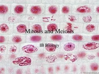
Microscopy is… • Using the microscope to enhance research techniques of cells at a microscopic level. • My project goal: The microscope was used to look at cells going through cell division (Mitosis).
Staining the Cells • Averaged around 8 hours • Used basic laboratory skills • Protocol varied depending on the cells
Background on Mitosis • Mitosis in single-celled organisms is responsible for the production of new individuals (asexual reproduction) • Single-celled organisms include Paramecia and Amoeba
Mitosis Mitosis in multi-cellular organisms is responsible for growth of the organismand repair of damaged tissues. Multi-cellular organisms include plants and animals
Genetic Material of the Cell • DNA (deoxyribonucleic acid) • 1. First it must be replicated so that there is a duplicate set of genetic information to be given to each daughter cell. • 2. Second, the genetic material (DNA) must be divided so that each daughter cell gets the exact same set of information.
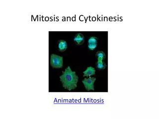
Mitosis is a 3 step process: 1. Replicationof genetic material in the mother cell 2.Separation of the replicated genetic material 3.Formation of the two daughter cells
Prophase Prophase: 1. Chromosomes condense 2. Nucleolus (or nucleoli) disappears 3. Nuclear membrane disappears 4.Spindle apparatus, composed of spindle fibers, forms and centrioles migrate apart. 5. Each chromosome becomes attached to a spindle fiber.
Metaphase Metaphase: 1. Chromosomes align in the "middle" (equator) of the cell. The chromosomes are pulled by the spindle fibers.
Anaphase Anaphase: 1. Centromere of each chromosome splits and one chromatid from each chromosome moves to centrioles at the poles of the cell. • The chromatids, which are now separate, are now called chromosomes. • There are now twice as many chromosomes in the cell as there were in the parent cell.
Telophase • Nuclear membranes reform around each group of newly divided chromosomes. • Nucleolus (or nucleoli) reappears • Spindles disappear • Chromosomes extend, becoming invisible • Cytokenesis occurs = cytoplasmic division of all the other materials in the cell (cytoplasm, cell membrane, organelles) that results in the formation of two new daughter cells with the correct number of chromosomes
Special Thanks To: Mentor: Mr. Mike Davidson In the Lab: John Griffin Nathan Claxton Dita Ishmaku
CIRL Staff Dr. Patricia Dixon Ms. Gina LaFrazza Ms. Stacy Vanderlaan Mr. Dave Sheaffer Mr. Carlos Villa
Finally Thank You to the… The National High Magnetic Field Laboratory and The National Science Foundation