Download
1 / 21
210 likes | 364 Views
Laser-induced Resonance Light Scatting Method for Detecting Trace Amounts of Curcumin in Solution. Chelsea Massaro, 12th grade Christian Home & Bible School. A.P. Biology Instructor: Ms. Krogel.

E N D
Laser-induced Resonance Light Scatting Method for Detecting Trace Amounts of Curcumin in Solution Chelsea Massaro, 12th grade Christian Home & Bible School A.P. Biology Instructor: Ms. Krogel I have participated in science fair for the past seven years. I have received Best of Show at my regional fair for six years. I have attended and placed at the state science fair for all seven years. I have attended the International Science and Engineering fair for three years, and qualified for the four year. I placed 4th at the International level in 2010. I have been working with Tandy Grubbs at Stetson University for four years. I have taken Chemistry, Biology, Anatomy and Physiology, Physical Science, A.P Environmental Science. I am currently take A.P. Biology. I plan on attending college at Florida State University and majoring in Analytical Chemistry.
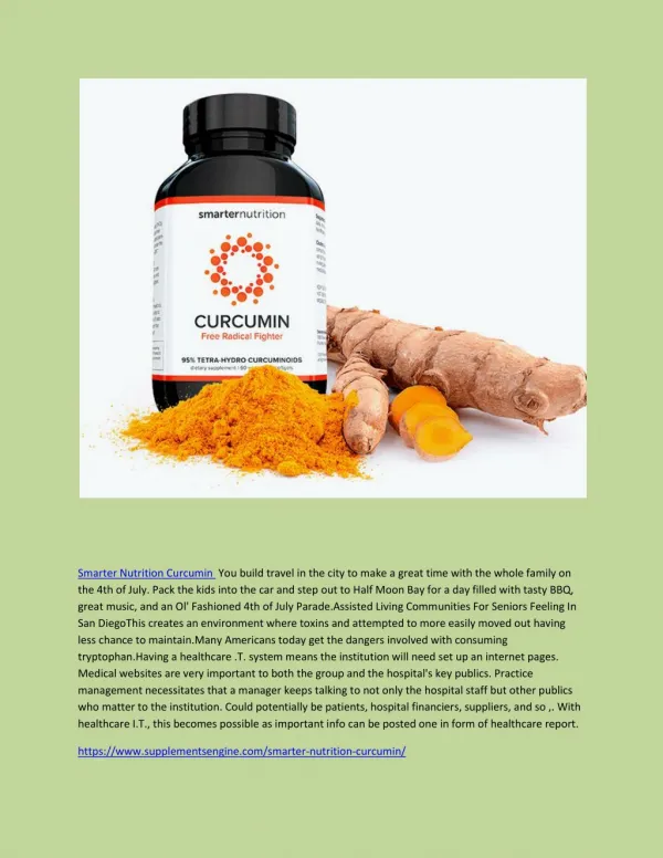
Introduction Curcumin is a curcuminoid found in the spice turmeric, which is often used in curries. Many experiments have showed that curcumin may have numerous medical qualities, although few have been confirmed. Like most herbal remedies, it was first used as a spice, and it was later discovered to have healing properties. Curcumin interests medical researchers because it shows many properties that may fight specific forms of cancer and Alzheimer’s disease. For my science fair project I chose to test if laser-induced resonance light scattering can be used to detect trace amounts of curcumin, and, if so, will the laser-induced be more precise than the fluorescence method. This subject interests me because I have an interest in chemistry. In addition, I have been researching uses of curcumin for four years and have become very interested in its numerous abilities. I have previously researched the use of curcumin as a chelating agent and discovered that the spice does have the ability to bind with certain transition metal ions. I have also researched curcumin’s ability to reduce the amount of oxidation that occurs to N-acetyl-tyrosine in an in vitro Cu(II)/H2O2 model of Alzheimer’s disease. I have also researched its ability to work as an antioxidant. I contacted Dr. Tandy Grubbs, professor of chemistry at Stetson University, and asked if he would work with me on this project.
Problem Can laser-induced resonance light scattering be used to detect trace amounts of curcumin in solution? If so, will the laser-induced method provide more precision than what is commonly available when a fluorescence spectrometer is used to measure the resonance light scattering? Hypothesis If a 150 milliwatt solid-state diode laser that emits at 532 nanometer is used to induce resonance light scattering in solution containing curcumin and copper (II), then trace amounts of curcumin will be detected in solution. If the laser-induced method is compared to the conventional fluorescence spectrometer based method of measuring resonance light scattering, then the laser-induced method will be more precise. Lab and stock solutions
Initial Research Alzheimer’s disease is a progressive neurodegenerative disease that is characterized by cognitive deterioration and neuron death, which causes memory impairment and disturbances in reasoning, language, perception, and planning. Alois Alzheimer, German psychiatrist, first described Alzheimer’s disease in 1906. He was studying slides prepared from the brain of a fifty-one year old woman who had suffered dementia and other symptoms that did not fit the definition of any known brain disorder at that time. The plaques and neurofibrillary tangles that are now used to identify Alzheimer’s disease during an autopsy were seen by Alzheimer in the slides. Alzheimer’s disease is the seventh-leading cause of death in the United States today. Free radicals, more specifically reactive oxygen species (ROS), are suspected to majorly contribute to Alzheimer’s disease. Free radicals are often formed when a weak bond between atoms splits, leaving an electron unpaired. This causes the atom to steal the needed electrons from surrounding atoms. ROS are derived from an oxygen molecule. Normally, they are formed in mitochondria and are highly unstable. In addition, ROS are able to damage macromolecules such as lipids, proteins, and nucleic acids in the body. Free radicals in the body can be extremely dangerous. They cause damage to DNA and cause cells to function poorly and deteriorate. The body normally uses antioxidants to protect against free radical damage. Damage takes place when free radical formation is excessive. Free radicals often cause aging, tissue damage, and diseases. They are suspected to have rolls in the development of cancer, strokes, and various heart diseases. Nerve cells in the brain are referred to as neurons. Neurons communicate and form networks. These networks carry signals and allow a person to multitask. Each network has a different job such as thinking, learning, remembering, hearing, smelling, and moving. In Alzheimer’s disease, neurons deteriorate and die causing the networks to fail, which causes confusion, disorientation, and memory loss. Senile plaques and neurofibrillary tangles are thought to have a major contribution to the neuron degeneration in Alzheimer’s disease. Senile plaques are abnormal deposits of protein and dead neurons. The plaques are normally found between neurons. They are made up of the protein fragment beta amyloid, which is produced by the body. Beta amyloid is a fragment of the protein amyloid precursor. Senile plaques interfere with communication in neurons. In a healthy brain, the fragments break down. In the brain of an Alzheimer’s disease patient, however, the fragments accumulate to form plaques.
Neurofibrillarytangles are twisted fibers often found inside neurons. The tangles mainly consist of the tau protein, which is found in the microtubule. The microtubule helps transport nutrients and other important substances from one part of the nerve cell to another. The tau protein stabilizes the microtubules. In Alzheimer’s disease, the tau protein is abnormal. This causes the microtubule structures to collapse thus forming tangles. Scientists are not absolutely sure what role senile plaques and neurofibrillary tangles play in Alzheimer’s disease. However, their formation is thought to block communication between neurons and disrupt activities neurons need to survive. In the Alzheimer’s disease patient’s brain, there is an overall shrinkage of brain tissue. The sulci, also referred to as grooves, in the brain widen. In addition, the well-developed folds, called the gyri, of the brain’s outer layer shrink. The ventricles also enlarge. The cerebral cortex and hippocampus often shrinks as well. The hippocampus is the area of the brain that controls memory. The cerebral cortex is the area of the brain that regulates thinking and behavior, as well as memory. The damaged nerve cells in the cerebral cortex and hippocampus often cause personality change and memory loss. Alzheimer’s disease leaves a patient with memory impairment, problems with communication, and personality and behavior changes. Curcumin is a yellow spice extracted from the dried roots of the Curcuma longa plant, which is part of the ginger family of herbs. Curcuma longa is native to southern and southeastern Asia. The spice has been studied extensively in medicine for the treatment of diseases such as cystic fibrous, hemorrhoids, various types of cancer, liver diseases, and arthritis. Curcumin also has a potential role in the treatment of Alzheimer’s disease (AD). ) (Mishra & Palanivelu, 2008) Although scientists are not sure of the cause of AD, there is evidence to suggest that oxidative stress, free radicals, and beta amyloid accumulation caused by metal toxicity contribute to Alzheimer’s disease pathology. (Balasubramanian, 2006) Cognitive functions in patients with Alzheimer’s disease could be improved by the use of curcumin as a chelation agent. (Mishra & Palanivelu, 2008) Studies show that metals ions could cause the beta amyloid accumulation causing the plaques found in a patient with AD. Metals such as copper have been revealed to induce the beta amyloid aggregation and increase the level of reactive oxygen species. (Balasubramanian, 2006) Curcumin, working as a chelation agent, would be able to prevent the neurotoxicity caused by metals. A study performed at the Chinese University of Hong Kong revealed that curcumin binds more effectively with redox-active metals such as copper and iron. (Mishra & Palanivelu, 2008) I have previously researched curcumin’s ability to bind with copper and discovered that it does work as an effective chelation agent. Further research indicated that the binding or curcumin to copper was occurring at the diketone group of the curcumin molecule.
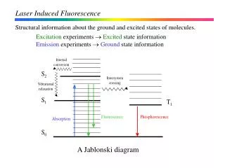
Resonance light scattering (RLS) is the process where light is forced to deviate from its original trajectory by the medium through which the light passes. Recently additional applications using RLS have been found. This includes the use of RLS to determine the micro-amount of biomolecules, metal ions, and other substances in solution. The RLS technique has become a highly sensitive analytical method. A study was previously done with RLS for the purpose of measuring amounts of curcumin in solution. When the RLS signal was taken of Cu(II) alone, it was relatively weak. However, when curcumin was added to a solution containing Cu(II) the signal was greatly enhanced. The chelation of curcumin to Cu(II) causes the amount of RLS to increase. Additionally, the enhanced RLS signal is proportional to the concentration of curcumin in the solution. The data in the study was taken using a Fluorescence spectrometer. The RLS signal was obtained by simultaneously scanning the excitation and emission monochromators from 300.0 to 700.0 nm. The excitation and emission silt widths were 5.0 nm. According to the results, the RLS signal was strongest at approximately 538 nm. (Chen, Chen, Guo, Song, & Zhu, 2008) Solid-state diode lasers are based on a solid-state gain medium, such as crystal or glass. A grain medium amplifies the light of laser beam. These lasers can emit light at 650 nm, 532 nm, and 405 nm. (Chen, Chen, Guo, Song, & Zhu, 2008) A laser-induced fluorescence method may produce the same results as the method previously mentioned. In laser-induced fluorescence, a solid-state diode laser could be used to excite a sample. When the sample’s electrons are excited, the energy level increases. However, this excitation lasts for only a few seconds. As the electrons lose energy, light is emitted. They fluoresce at a longer wavelength than the wavelength of the laser. Due to unique energy states in each atom, the fluorescence, or resonance light scattering, could be used for the identification of a substance in solution. (Harris, 2008) Experimental solutions
Work Cited Abramowitz, M., & Davidson, M.W. (n.d.) Concepts of Digital Imaging Technology: Photomultiplier Tubes. Retrieved from http://learn.hamamatsu.com/articles/photomultipliers.html Balasubramanian, K. (2006, April 20). Molecular Orbital Basis for Yellow Curry Spice Curcumin's Prevention of Alzheimer's Disease. Journal of Agriculture and Food Chemistry, 54(10), 3512-3520. Dio: 10.1021/jf0603533 Baum, L., Ng, A. (2004). Curcumin interaction with copper and iron suggests one possible mechanism of action in Alzheimer's disease animal models. Journal of Alzheimer's Disease, 6(4,) 367-377. Retrieved from http://iospress.metapress.com/content/yy1rf8ctbf08lulu/ Chen, J., Chen, Z., Guo, Z., Song, T., Zhu, L. (2008). A novel curcumin assay with the metal ion Cu (II) as a simple probe by resonance light scattering technique. Elsevier, (72), 518-522. Harris, W. (2008). How Laser Analysis Works. Retrieved from http://science.howstuffworks.com/laser- analysis3.htm Hidaka, K., Maekawa, T., Masuda, T., Shinohara, A., Takeda, Y., & Yamaguchi H. (1999).Chemical Studies on Antioxidant Mechanism of Curcuminoid: Analysis of Radical Reaction Products from Curcumin. Journal of Agricultural and Food Chemistry, 47(1), 71-77. doi: 10.1021/jf9805348 Laser. (2012). In Encyclopedia Britannica online. Retrieved from http://www.britannica.com/EBchecked/topic/553349/solid-state-diode-laser Mishra, S., & Palanivelu, K. (2008). The effect of curcumin(turmeric) on Alzheimer’s Disease: An overview. Official Journal of Indian Academy of Neurology, 11(1), 13-19. dio: 10.4103/0972-2327.40220 N.A. (2009). Focal Length. Retrieved from http://amazing-space.stsci.edu/resources /explorations/groundup/lesson/glossary/term-full.php?t=focal_length Scott, R. (2008). Fluorescence Spectrometer. Retrieved from http://www.chromatography- online.org/topics/fluorescence/spectrometer.html
Materials • .1 mL volumetric pipette • .01 mL volumetric pipette • Bulb pipette • Four 100 mL flasks • 125 mL flask • Eight 10 mL flasks • Cuvette • Kimtechkimwipes • pH meter and probe • Pen or pencil and Log book • Labels • Plastic garbage bag • lint free paper towels • Rubber gloves, lab coat, • Laser goggles and eye protection • 150 milliwatt Solid-state Diode Laser • Fluorescence Spectrometer • Weight boats • Analytic balance • Spatula • Sodium phosphate monobasic monohydrate • Curcumin • Sodium hydroxide solution • Copper (II) sulfate pentahydrate • Distilled water • Ethyl alcohol • 1 mL volumetric pipette Independent Variables Dependent Variables • Amount of curcumin added to experimental solution • Amount of resonance light scattering Constants • Concentrations of stock solutions • Total volume in each experimental sample • Amount of sodium phosphate monobasic monohydrate buffer stock solution and copper (II) stock solution in each experimental sample • Set up of solid-state diode laser • Fluorescence spectrometer settings
Control Group • Solution containing 1 mL phosphate monobasic monohydrate buffer stock solution, 4 mL Cu(II) stock solution, and 5 mL of the dilution solution (50 mL ethyl alcohol and 50 mL distilled water) Experimental Group • Solution containing 1 mL phosphate monobasic monohydrate buffer stock solution, 4 mL Cu(II) stock solution, 1 mL curcumin stock solution, and 4 mL of the dilution solution (50 mL ethyl alcohol and 50 mL distilled water) • 1 mL phosphate monobasic monohydrate buffer stock solution, 4 mL Cu(II) stock solution, .5 mL curcumin stock solution, and 4.5 mL of the dilution solution (50 mL ethyl alcohol and 50 mL distilled water) • 1 mL phosphate monobasic monohydrate buffer stock solution, 4 mL Cu(II) stock solution, .2 mL curcumin stock solution, and 4.8 mL of the dilution solution (50 mL ethyl alcohol and 50 mL distilled water) • 1 mL phosphate monobasic monohydrate buffer stock solution, 4 mL Cu(II) stock solution, .1 mL curcumin stock solution, and 4.9 mL of the dilution solution (50 mL ethyl alcohol and 50 mL distilled water) • 1 mL phosphate monobasic monohydrate buffer stock solution, 4 mL Cu(II) stock solution, .05 mL curcumin stock solution, and 4.95 mL of the dilution solution (50 mL ethyl alcohol and 50 mL distilled water) • 1 mL phosphate monobasic monohydrate buffer stock solution, 4 mL Cu(II) stock solution, .02 mL curcumin stock solution, and 4.98 mL of the dilution solution (50 mL ethyl alcohol and 50 mL distilled water) • 1 mL phosphate monobasic monohydrate buffer stock solution, 4 mL Cu(II) stock solution, .01 mL curcumin stock solution, and 4.99 mL of the dilution solution (50 mL ethyl alcohol and 50 mL distilled water)
Procedure • Perform all steps under the supervision of the Qualified Scientist in University laboratory. • Put on disposable rubber gloves, lab coat, and eye protection. Have the designated supervisor do so as well. • Preparation of sodium phosphate monobasic monohydrate buffer stock solution: • Using an analytic balance, spatula, and weight boat, measure 4 grams of sodium phosphate monobasic monohydrate buffer into a 125 mL flask. • Add distilled water into the flask containing the sodium phosphate monobasic monohydrate buffer to obtain a total volume of 75 mL. • Using a bulb pipette, add aliquots of one molar sodium hydroxide solution until reaching a pH of 7.4. Use a pH meter and probe to monitor the pH of the solution. Dilute with distilled water until obtaining a volume of 125 mL. Mix well. • Clean surface analytic balance using KimtechKimwipes. Clean spatula with distilled water and dispose of weight boat in trash. • Preparation of Cu(II) Stock Solution: • Using an analytic balance, spatula, and weight boat, measure .25 grams of Cu(II) into a 100 mL flask. • Add sodium phosphate monobasic monohydrate buffer stock solution into flask containing Cu(II) to obtain a total volume of 100 mL. Mix well. • Using a volumetric pipette, place 1 mL of the above solution into a separate 100 mL volumetric flask. Add distilled water until obtaining a volume of 100 mL. • Clean surface analytic balance using KimtechKimwipes. Clean spatula with distilled water and dispose of weight boat in trash. • Preparation of Curcumin Stock Solution: • Using an analytic balance, spatula, and weight boat, measure .004 grams of curcumin into a 100 mL flask. • Add 50 mL ethyl alcohol and 50 mL distilled water to flask containing curcumin to obtain a total volume of 100 mL. Mix well. • Clean surface analytic balance using KimtechKimwipes. Clean spatula with distilled water and dispose of weight boat in trash. • Add 50 mL distilled water and 50 mL ethyl alcohol to a 100 mL flask to create dilution solution.
Using a 1 mL volumetric pipette, add 1 mL phosphate monobasic monohydrate buffer stock solution, 4 mL Cu(II) stock solution, 1 mL curcumin stock solution, and 4 mL of the dilution solution into a 10 mL flask. Label flask as ‘11 μM Curcumin Sample’. • Using a 1 mL volumetric pipette, add 1 mL phosphate monobasic monohydrate buffer stock solution, 4 mL Cu(II) stock solution, .5 mL curcumin stock solution, and 4.5 mL of the dilution solution into a 10 mL flask. Label flask as ‘5.5 μM Curcumin Sample’. • Using a 1 mL volumetric pipette, add 1 mL phosphate monobasic monohydrate buffer stock solution, 4 mL Cu(II) stock solution, and 4.8 mL of the dilution solution into a 10 mL flask. Using .1 mL volumetric pipette, add .2 mL curcumin stock solution to the same flask. Label flask as ‘2.2 μM Curcumin Sample’. • Using a 1 mL volumetric pipette, add 1 mL phosphate monobasic monohydrate buffer stock solution, 4 mL Cu(II) stock solution, and 4.9 mL of the dilution solution into a 10 mL flask. Using .1 mL volumetric pipette, add .1 mL curcumin stock solution to the same flask. Label flask as ‘1.1 μM Curcumin Sample’. • Using a 1 mL volumetric pipette, add 1 mL phosphate monobasic monohydrate buffer stock solution, 4 mL Cu(II) stock solution, and 4.95 mL of the dilution solution into a 10 mL flask. Using .1 mL volumetric pipette, add .05 mL curcumin stock solution to the same flask. Label flask as ‘.55 μM Curcumin Sample’. • Using a 1 mL volumetric pipette, add 1 mL phosphate monobasic monohydrate buffer stock solution, 4 mL Cu(II) stock solution, and 4.98 mL of the dilution solution into a 10 mL flask. Using .01 mL volumetric pipette, add .02 mL curcumin stock solution to the same flask. Label flask as ‘.22 μM Curcumin Sample’. • Using a 1 mL volumetric pipette, add 1 mL phosphate monobasic monohydrate buffer stock solution, 4 mL Cu(II) stock solution, .01 mL curcumin stock solution, and 4.99 mL of the dilution solution into a 10 mL flask. Using .01 mL volumetric pipette, add .01 mL curcumin stock solution to the same flask. Label flask as ‘.11 μM Curcumin Sample’. • Using a 1 mL volumetric pipette, add 1 mL phosphate monobasic monohydrate buffer stock solution, 4 mL Cu(II) stock solution, and 5 mL of the dilution solution into a 10 mL flask. Label flask as ‘Control Sample’. • See Diagram A for solid-state diode laser set up. • Remove eye protection and put on laser goggles. Have the designated supervisor do so as well. • Turn on solid-state diode laser. Allow it to warm up for 10 minutes. • Turn on photomultiplier tube set to 750 volts. • Using a 1 mL volumetric pipette, insert 2 mL of sample into a cuvette. • Place cuvette into laser set up. • Record results from photomultiplier tub for sixty seconds. • Remove cuvette from laser set up and dispose of solution as indicated in step 42. • Dispose of solution in flask as indicated in step 42.
Wash cuvette with warm, soapy water. Rinse and dry cuvette. • Repeat steps 23 - 28 for each sample created in steps 10 - 17. • Turn on fluorescence spectrometer. The settings for the spectrometer are as follows: Excitation band width: 10, Emission band width: 20, Response time: 8 seconds, Sensitivity: High, Auto shutter: On • Repeat steps 10 - 17 to create samples. • Using a 1 mL volumetric pipette, insert 2 mL of distilled water into cuvette. Place cuvette in Fluorescence Spectrometer. Use this as the autozero. • Remove cuvette and wash it with warm, soapy water. Rinse and dry cuvette. • Using a 1 mL volumetric pipette, insert 2 mL of sample into cuvette. • Place cuvette into Fluorescence Spectrometer. • Run Fluorescence Spectrometer. Record results. • Remove cuvette. • Dispose of solution in cuvette and flask as indicated in step 42. • Wash cuvette with warm, soapy water. Rinse and dry cuvette. • Repeat steps 33 - 38 for each sample. • Wash all used glassware with warm, soapy water. Rinse and dry. • Dispose of all stock solutions and samples as indicated in step 42. • All solutions will be disposed of by the lab manager of Stetson University using the following procedure. • The transition metal ion solutions will be disposed of by the lab manager of Stetson University using the following procedure Pour the transition metal ion solution into a large dish • Allow the water to slowly evaporate out of the dish • After the water has fully evaporated, dissolved components are left behind as a solid. Stetson University accumulates the components and combines them with a large amount of mixed transition metal salts gathered over the course of a year. • After a year, Stetson University sends the salts to a waste disposal company which properly disposes of the waste. • Dispose all paper waste in a sealed plastic garbage bag and place in a garbage can for pick up by disposal service. • Return all other lab equipment to laboratory storage • Return all unused chemicals to chemical storage at University • Repeat all steps for each trial.
Solid-State Diode Laser Set-up Photo Diode Light Detector Photo- multiplier Tube Set to 750 V Focal Length 5 cm Iris Monochromator Solid-State Diode Laser Set-up Photo Diode Light Detector Sample Exit Slit Entrance Slit Collection Lens Photo- multiplier Tube Set to 750 V Focal Length 5 cm Iris Monochromator Solid-State Diode LaserExcitation at 532 nanometers Sample Mirror Exit Slit Entrance Slit Collection Lens Solid-State Diode LaserExcitation at 532 nanometers Mirror Solid-State Diode Laser Set-up Photo Diode Light Detector Photo- multiplier Tube Set to 750 V Focal Length 5 cm Iris Monochromator Sample Exit Slit Entrance Slit Collection Lens Solid-State Diode LaserExcitation at 532 nanometers Mirror
Safety Measures • Work in a University Chemistry Laboratory • Work under the supervision of qualified scientist. • Wear a lab coat, disposable gloves, laser goggles, and eye protection. • Avoid inhalation, eye contact, skin contact, and ingestion of the nitrates and curcumin • Keep chemicals and solutions away from heat, sparks, and flame • Wash hands thoroughly after experimentation Clean up and Waste Disposal • Paper waste will be sealed in a plastic garbage bag and placed in garbage can for pick up by disposal service. • Return all other lab equipment to laboratory storage. • Return all unused chemicals to chemical storage at University. • Using hot water, rinse the curcumin stock solution down the drain of a sink. • All other solutions will be disposed of by the lab manager of Stetson • University using the following procedure: • Pour the transition metal ion solutions collected in the waste jar into a large dish. • Allow the water to slowly evaporate out of the dish. • After the water has fully evaporated, dissolved components are left behind as a solid. Stetson University accumulates the components and combines them with a large amount of mixed transition metal salts gathered over the course of a year. • After a year, Stetson University sends the salts to a waste disposal company which properly disposes of the waste.
Data Analysis • Calculate the average resonance light scattering in each solution for each method (laser-induced and fluorescence spectroscopy). • Place data in an x-axis, y-axis plot. • Insert the linear trend line. • Method is viable if average results varied only slightly from linear trend line. • Calculate the standard deviation of the results of all trials for each method (laser-induced and fluorescence spectroscopy). • Compare the standard deviation of the laser-induced method to the fluorescence spectroscopy method to determine which method is more precise. • A lower standard deviation indicts a more precise method. Resonance light scattering of experimental solution
Discussion A slight curvature was observed in the data. I believe this was due to the curcumin in the solution precipated out. Curcumin is not soluble in water. Each experimental solution was diluted with a solution containing 50% ethanol and 50 % distilled water. When the curcumin begins to precipate out of the solution, due to the percentage of water to ethanol, the resonance light scattering signal was disturbed. This caused the slight curvature. Future experiments will be performed using a higher percentage of ethanol and a lower percentage of distilled water. Conclusion The key results for this experiment are the resonance light scattering results from both the solid-state diode method and the fluorescence spectrometer method. Both sets of data were shown to be linear, indicating that the light scattering intensity increased linearly with increasing curcumin concentration. The standard deviation of the measured light scattering intensity was calculated after performing multiple trials. Formation of complexes can be detected with high precision by laser-induced resonance light scattering at 532 nanometers. Results indicated that the intensity of the resonance light scattering signal is proportional to the amount of curcumin in solution. The standard deviation results indicated that the solid-state laser method is the more precise method, supporting the hypothesis. Additionally, laser-induced resonance light scattering was capable of detecting curcumin at concentrations as low as 10 nanograms per milliliter.
Value of Experiment The value of this experiment is that due to the large of amount of resonance light scattering given off by the binding of curcumin to copper, a 150 milliwatt solid-state diode laser can be used to detect trace amounts of curcumin in solution. The laser-induced resonance light scattering method investigated here could be helpful in precisely measuring curcumin concentrations present in a blood sample if curcumin was used as treatment for disease. Acknowledgements I would like to thank Dr. Tandy Grubbs, Professor of Chemistry at Stetson University for helping me with my project. He assisted me in determining which metals to test and the most accurate method of testing. In addition, Dr. Grubbs showed me how to conduct the experiment. Without his advice and assistance this project would have never happened. I would also like to thank Stetson University for allowing me to conduct my experiments in one of their labs and to use their materials.
Bibliography Copper Nitrate MSDS (2011). Copper Nitrate. Retrieved from http://www.palomar.edu/ehs/Chemistry%20MSDS/COPPER% 20NITRATE.pdf Curcumin MSDS (2010). Curcumin. Retrieved from http://www.palomar.edu/ehs/Chemistry%20MSDS/TUMERIC.pdf Ethyl Alcohol MSDS (2010). Ethanolfromhttp://www.sigmaaldrich.com/catalog/ProductDetail.do?lang=en&N4=459836|SIAL&N5=SEARCH_CONCAT_PNO|BRAND_KEY&F=SPEC HydrogenPeroxide MSDS (2009). HydrogenPeroxide. Retrieved from http://www.sigmaaldrich.com /catalog/ProductDetail.do?lang=en&N4=316989|SIAL&N5=SEARCH_CONCAT_PNO|BRAND_KEY&F=SPEC International Rules-For Precollege Science Research: Guidelines for Science and Engineering Fairs (2011). ISEF. Retrieved from http://www.societyforscience.org/document.doc?id=9 Phosphate Buffer MSDS (2009). Phosphate Buffer. Retrieved from http://www.sigmaaldrich.com /catalog/ProductDetail.do?lang=en&N4=P3288|SIGMA&N5=SEARCH_CONCAT_PNO|BRAND_KEY&F=SPEC Sodium Hydroxsol MSDS(2010). Sodium Hydroxsol. Retrieved from http://www.fishersci.com/msds?productName =SS2761&productDescription=SODIUM+HYDROX+SOL+N%2F10+CR+1L&catNo=SS276- 1&vendorId=VN00033897&storeId=10652 Solid-state diode laser set-up
Oxygen – red Carbon – blue Hydrogen – white Copper (II) – green Curcumin Binding to Copper(II) Solid-state diode laser Mixing experimental solutions All photos by T. Massaro