Download
1 / 46
680 likes | 906 Views
ANATOMY OF THE NERVOUS SYSTEM. Lecture 9 &10. Functions of the nervous system. 1. Initiate and/or regulate movement of body parts 2. Regulate secretions from glands 3. Gather information about the external environment and the internal environment of the body

E N D
ANATOMY OF THE NERVOUS SYSTEM Lecture 9 &10
Functions of the nervous system • 1. Initiate and/or regulate movement of body parts • 2. Regulate secretions from glands • 3. Gather information about the external environment and the internal environment of the body • using senses (sight, hearing, touch, balance, taste) and mechanisms to detect pain, temperature, pressure, and certain chemicals, such as carbon dioxide, hydrogen, and oxygen • 4. Maintain an appropriate state of consciousness • 5. Stimulate thirst, hunger, fear, rage, and sexual behaviors appropriate for survival
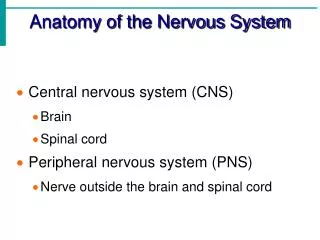
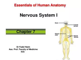
The entire nervous system can be divided into two parts: • 1- Central nervous system (CNS) • includes the brain and spinal cord • 2- Peripheral nervous system (PNS), which consists of: • Cranial nerves • spinal nerves
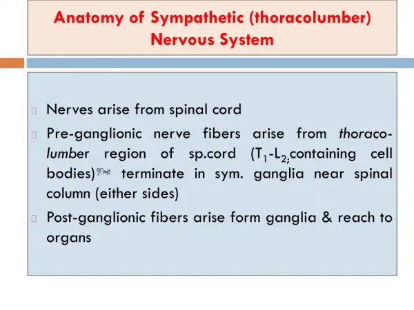
A further distinction is often made: • the autonomic nervous system (ANS) • which integrates activity of visceral structures • smooth muscle, cardiac muscle, and glands • The ANS has elements in both: • Central nervous system. • Peripheral nervous system.
In the PNS: 1- sensory (afferent) nerves - gather information about the external and internal environments and relay this information to the CNS. - The specialized organs or cells that detect specific stimuli are sensory receptors.
CNS interprets information arriving via the PNS, integrates that information, and initiates • appropriate movement of body parts • glandular secretion • behavior response. 2- Motor efferent - Communication between the CNS and muscles and accomplished via nerves of the PNS
Microscopic Neuroanatomy • The individual nerve cell is called a neuron. • Each neuronal cell body gives rise to one or more nerve processes and cytoplasmic extensions of the cell. • The nerve processes are called dendritesif they transmit electrical signals toward the cell bodies • They are called axonsif they conduct electrical signals away from the cell bodies.
Neurons may be classified morphologically according to their number of nerve processes: • Unipolar neuronshave one process • Bipolar neuronshave one dendrite and one axon • Multipolar neuronshave a number of dendrites in addition to their single axon. • We do not have true unipolar neurons, but many sensory neurons have their single dendrite and axon fused so as to give the appearance of a single process • This configuration is pseudounipolar.
Nervous tissue consists not only of neurons but also of supportive cells. • In the CNS, these supportive cells are the neuroglia, comprising a variety of glial cells • Groups of nerve cell bodies within the CNS are generally callednuclei, while groups of nerve cell bodies in the PNS are calledganglia. • In general terms: • Aggregates of neuronal cell bodies form the gray matterof the CNS • Regions characterized primarily by tracts arewhite matter.
Development of CNS • Shortly after gastrulation, ectodermal cells on the dorsum just cranial to the primitive streak begin to proliferate and differentiate into a neural plate. • The neural plate proliferates faster along its lateral margins than on the midline, creating the neural groove
Development of CNS • The edges of which (the neural folds) ultimately meet dorsally to form the neural tube. • The entire CNS is formed from the cells of the neural tube. • The lumen of the neural tube persists in the adult as the central canal of the spinal cord and as the ventricles of the brain.
Development of the spinal cord continues by an increase in the thickness of the wall of the neural tube. • As cells divide and differentiate, three concentric layers of the neural tube emerge: • an inner (ventricular zone) • a middle (intermediate zone) • a superficial (marginal zone)
The thin ventricular zone of cells (also called ependymal zone) surrounds the lumen of the neural tube • The site of mitosis of neuronal and glial precursors in the developing nervous system. • It will ultimately form the ependyma of the central canal of the spinal cord and of the ventricles of the brain.
As cells are born in the germinal layer, they migrate outward to form the intermediate zone(also called mantle zone). • The intermediate zone comprises neurons and neuroglia and becomes the gray matter near the center of the cord. • The dorsal parts of the intermediate zone develop into the dorsal horns. - The ventral intermediate zone becomes the ventral horns
The marginal zone, which is most superficial, consists of nerve processes that make up the white matter of the spinal cord. • The spinal cord white matter is divided into dorsal, lateral, and ventral funiculi, which are delimited by the dorsal and ventral horns of gray matter.
Development of the brain: • The first gross subdivisions of the brain create the three-vesicle stage. • These subdivisions, which consist of three dilations of the presumptive brain, are • prosencephalon, or forebrain • mesencephalon,or midbrain • rhombencephalon,or hindbrain. • In the five-vesicle stage of development • the prosencephalon further subdivides to form the telencephalon (future cerebrum) and the diencephalons , • the rhombencephalons divides into the Metencephalon (future pons and cerebellum) and the myelencephalon(future medulla oblongata). • The mesencephalon does not subdivide.
Central Nervous System • Brain • The gross subdivisions of the adult brain include: • cerebrum • cerebellum • brainstem • The cerebrum develops from the embryonicTelencephalon. • The components of the brainstem are defined in a number of ways • include the diencephalon, midbrain, pons, medulla oblongata
Cerebrum comprises: • the two cerebral hemispheres, including the cerebral cortex, the basal nuclei • The surface area of the cerebrum in domestic mammals is increased by numerous foldings to form convex ridges, called gyri (singular gyrus),which are separated by furrows called fissures or sulci. • A particularly prominent fissure, the longitudinal fissure, lies on the median plane and separates the cerebrum into its right and left hemispheres. • Deep to the cerebral cortex are aggregates of subcortical gray matter called the basal nuclei
Diencephalon • Is a derivative of the prosencephalon. • thalamus • epithalamus • hypothalamus • the third ventricle are included in the diencephalon. • The thalamusis an important relay center for nerve fibers connecting the cerebral hemispheres to the brainstem and spinal cord. • The epithalamus, dorsal to the thalamus, includes a number of structures, the pineal gland, which is an endocrine organ in mammals. • The hypothalamus, ventral to the thalamus, surrounds the ventral part of the third ventricle and comprises many nuclei that function in autonomic activities and behavior. • Attached to the ventral part of the hypothalamus is the hypophysis, or pituitary gland
Mesencephalon • The mesencephalon, or midbrain • lies between the diencephalon rostrally and the pons caudally. • The two cerebral peduncles • four colliculi are the most prominent features of the midbrain. • The cerebral peduncles, also called crura cerebrii, are large bundles of nerve fibers connecting the spinal cord and brainstem to the cerebral hemispheres. • These peduncles consist of both sensory and motor fiber tracts. • The colliculiare four small bumps (colliculusis Latin for little hill) on the dorsal side of the midbrain. • They consist of right and left rostral colliculi and right and left caudal colliculi. • The rostral colliculi coordinate certain visual reflexes, • The caudal colliculi are relay nuclei for audition (hearing).
Metencephalon. • The metencephalonincludes • the cerebellum dorsally and the pons ventrally. • The cerebellum features two lateral hemispheres and a median ridge called the vermis. • The surface of the cerebellum consists of many laminae called folia. In the cerebellum, like the cerebrum, the white matter is central, and the gray matter is peripheral in the cerebellar cortex. • The cerebellum is critical to the accurate timing and execution of movements; it acts to smooth and coordinate muscle activity. • The pons is ventral to the cerebellum, and its surface possesses visible transverse fibersthat form a bridge from one hemisphere of the cerebellum to the other.
Myelencephalon • The myelencephalonbecomes the medulla oblongatain the adult. • It is the cranial continuation of the spinal cord • The medulla oblongata (often simply called the medulla) • contains a number of important autonomic centers and nuclei for cranial nerves.
Ventricular System • The ventricles of the brain are the remnants of the lumen of the embryonic neural tube. • Right and left lateral ventricleslie within the respective cerebral hemispheres. • They communicate with the midlinethird ventricle by way of the interventricular foramina. • Most of the third ventricle is surrounded by the diencephalon. • The third ventricle connects with the fourth ventricle by way of the mesencephalic aqueduct(cerebral aqueduct) passing through the midbrain. • The fourth ventricle, between the cerebellum above and pons and medulla below, communicates with the subarachnoid spacesurrounding the CNS through two lateral apertures.
Each ventricle features a choroid plexus • a tuft of blood capillaries that protrudes into the lumen of the ventricle. • The plexus of capillaries is covered by a layer of ependymal cells that are continuous with the lining membrane of the ventricles. • Cerebrospinal fluid (CSF), filling the ventricular system and surrounding the CNS, is formed primarily by the choroid plexuses, with a smaller contribution made by the ependyma lining the ventricles. • CSF is a modified transudate, formed primarily through active secretion by the ependymal cells, especially those of the choroid plexuses.
Meninges • The coverings of the brain and spinal cord are the meninges (singular meninx). • They include, from deep to superficial • the pia mater • the arachnoid • the dura mater. • The pia mater, the deepest of the meninges, is a delicate membrane that invests the brain and spinal cord, following the grooves and depressions closely. • The pia mater forms a sheath around the blood vessels and follows them into the substance of the CNS.
Meninges • The arachnoid • arises embryologically from the same layer as the pia mater but separates from it during development so that a space forms between them. • Because of the weblike appearance of these filaments, this middle layer is called the arachnoid (arachnoidea, arachnoid mater). • Together, the pia mater and arachnoid constitute the Ieptomeninges (from the Latin word lepto, delicate), reflecting their fine, delicate nature. • The space between the two layers is the subarachnoid space. • It is filled with CSF.
Meninges • The Dura mater is the tough fibrous outer covering of the CNS. • Within the cranial cavity the dura mater is intimately attached to the inside of the cranial bones and so fulfills the role of periosteum. • It also forms the falx cerebri, a median sickle-shaped fold that lies in the longitudinal fissure and partially separates the cerebral hemispheres. • Another fold of dura mater, the tentorium cerebelli, runs transversely between the cerebellum and the cerebrum. • In some locations within the skull, the dura mater splits into two layers divided by channels filled with blood. These dural sinuses receive blood from the veins of the brain and empty into the jugular veins. • They are also the site of reabsorption of CSF back into the circulation.
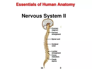
Spinal Cord • The spinal cord is the caudal continuation of the medulla oblongata. • Unlike that of the cerebrum, the spinal cord’s gray matter is found at the center of the cord, forming a butterfly shape on cross-section. • Myelinated fiber tracts, the white matter, surround this core of gray matter. • A spinal cordsegment is defined by the presence of a pair of spinal nerves. Spinal nerves are formed by the conjoining of dorsal and ventral roots.
Spinal Cord • Sensory neurons of neural crest origin are present in aggregates, called dorsal root ganglia, lateral to the spinal cord. • The neurons within these ganglia are pseudounipolar • they give rise to axons that enter the dorsal horn of the spinal cord and other fibers that join with motor fibers from the ventral horn neurons to become a spinal nerve extending into the periphery. • The ventral root of the spinal nerve consists largely of motor fibers that arise from the nerve cells in the ventral horn of the spinal cord. • The dorsal and ventral roots unite to form the spinal nerve close to the intervertebral foramen between adjacent vertebrae.