Download
1 / 43
430 likes | 438 Views
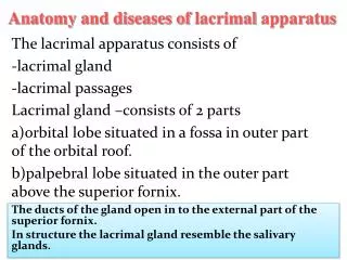
Orbit tumours (including lacrimal gland). CASE 1. Mass in supero-temporal orbit Painless proptosis. Lacrimal gland. Well defined, lesion. Neoplastic glands embedded in a fibro (pink) -myxoid (blue) matrix.

E N D
CASE 1 Mass in supero-temporal orbit Painless proptosis
Lacrimal gland Well defined, lesion
Neoplastic glands embedded in a fibro (pink) -myxoid (blue) matrix
Ducts are composed of bilayer of benign epithelium (inner epithelium and outer myoepithelial)
Benign pleomorphic adenoma Commonest benign lacrimal gland tumour. Orbital lobe usually-painless Frank bone invasion not present. Can become malignant Shouldn’t really biopsy-can seed and recur. Psuedo-encapsulated. Can transform into carcinoma.
CASE 2 Painful superotemporal orbital mass.
Swiss cheese appearance. Smoothly rounded sheets of deceptively Bland basaloid cells, containing round pools of mucin that mimic glands
Tumour on both sides of pink lacrimal gland capsule = infiltration=malignant
Adenoid cystic carcinoma 2nd most common epithelial lacrimal gland tumour Malignant Perineural invasion-therefore painful Various histological patterns Adenoid-’gland like’ bacause not true glands. The mucin pools develop after stroma is pinched off from outside. Poor prognosis
For Lymphoma (Maltoma) See conjunctival session-exactly the same appearance
HISTORY • CHILD WITH PROPTOSIS
Spindle cells in loose matrix, with scattered deep pink larger cells.
Cells positive for Muscle marker Desmin.
Tumour nuclei positive for Muscle transcription factor MyoD1
Rhabdomyosarcoma(embryonal type) Sarcoma Arise from pluripotential mesenchymal cells Commonest malignant orbital tumour of childhood Muscle marker positive Electron microscopy shows striations (can see on light microscopy sometimes) and Z bands. Immunohistochemistry detects muscle proteins-Desmin and MyOD1 transcription factor. After biopsy-chemo and radiotherapy usually 95 % 5 y survival for embryonal subtype.
Other one…..Alveolar rhabdomyosarcoma • Nests of smaller round blue cells, with central discohesion. Peppered with some larger cells with pink cytoplasm. • Has 1: 13 or 2:13 specific and diagnostic cytogenetic translocation • Worse prognosis than embryonal
A note on orbital tumours • Adult-commonest primary-non-Hodgkin’s lymphoma. • Kids-commonest primary-rhabdomyosarcoma. • Other tumours in orbit: Adults-Primary orbital tumours: liposarcoma, malignant peripheral nerve sheath tumour, solitary fibrous tumour. Remember metastatic carcinoma. • Children-primary alveolar soft part sarcoma. Metastatic tumours commoner: neuroblastoma, leukaemia, Ewing’s sarcoma, Wilm’s tumour are commoner ones (Jerry and Carol Shields series).
HISTORY • 8 YEAR-OLD CHILD • REDUCED UNILATERAL VISION • FUSIFORM SWELLING OF OPTIC NERVE
ABNORMAL OPTIC NERVE expansion
Expansion by tumour showing process bearing cells
Process bearing cells alternating with fibrous areas containing intense pink bodies called Rosenthal fibres
Optic nerve pilocytic astrocytoma Unilateral visual loss and proptosis First 2 decades of life. NF-1 association Fusiform swelling of ON Confined by dura Extends through optic foraman to chaism. Cells, with many long delicate fibres, with more fibrous areas containing Rosenthal fibres. Cystic areas. Tumour often involves subarachnoid space-cardinal features. Rosenthal fibre = collections of alpha-beta crystalline. Slowly growing tumour-hardly ever fatal.
HISTORY • 40 YEAR OLD WOMAN • UNILATERAL VISUAL LOSS • PROPTOSIS • IMAGING SHOWS MASS AT SPHENOID RIDGE
Tumour composed of bland spindly cells With occasional foci of dystrophic calcification
Bland Interlacing spindle cells Bland=do not look cytologically malignant
Meningioma Mostly benign Optic nerve sheath OR Sphenoid wing origins Many patterns-but classicial is whorls of meningothelial cells, +/-psammoma bodies (calcification arranged as concentric circles-like rings of a tree) Arise from arachnoid cells Female bias Express progesterone receptors Associated with NF-2