Download
1 / 40
400 likes | 407 Views
Explore the field of tissue engineering and regenerative medicine, which aims to restore damaged tissues through the use of cells, hormones, and support scaffolds. This article covers the principles, techniques, and future prospects of tissue engineering.

E N D
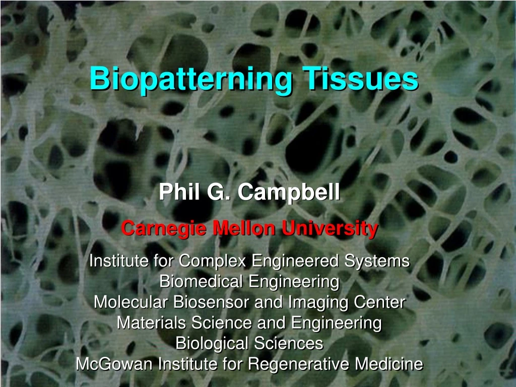
Biopatterning Tissues Phil G. Campbell Carnegie Mellon University Institute for Complex Engineered Systems Biomedical Engineering Molecular Biosensor and Imaging Center Materials Science and Engineering Biological Sciences McGowan Institute for Regenerative Medicine
What is Tissue Engineering/Regenerative Medicine? Seeks to understand the basic biology behind tissue regeneration and apply those concepts to restore structure and function of damaged tissues. Approaches include the application of cells, hormones and/or support scaffolds directly in vivo or in vitro for future in vivo implantation or as extracorporeal devices.
The Ultimate Vision for Tissue Engineering: Complete Tissue Regeneration Spinal Cord Upper and Lower Jaw Retina and Lens Tail Heart Over 20 days Limb The Newt Adapted from Brockes From Dr. Susan Bryant, Univ. of Calif., Irvine
Principles of Tissue Engineering Cells ECM Defect Regeneration Hormones Blood Supply
What a medical robotic bioprinting system might look like in the near/far future. Source: The Fifth Element (1997)
Solid-Phase (Immobilized) Hormone Patterns May be Relevant wt/wt VEGF with heparin binding domain 120/120 VEGF without heparin binding domain Ruhrberg, C., et al. Genes and Development 2002, 16:2684-2698
Printing Biology (Bioprinting) What are Bioinks? What are Biopapers? Focus on Tissue Engineering – Repair or replacement of dysfunctional tissues
2D and 3D BioPrinters 2D Printer 3D Printer Growth factor directed cell response In situ printing scaffolds/growth factors Weiss, L.E., et al. Computer-Aided Design 2005;37:1127-1139. Campbell, P. G., et al. Biomaterials 2005;26:6762-6770.
Piezoelectric inkjet Modulating Surface Concentrations with Inkjet Printing 30 mm nozzle 14 ± 4 pl drop Concentration gradient of 605 nm emission streptavidin-conjugate quantum dots (sAvQdots) printed on a nitrocellulose-coated glass slide
Combinatorial Arrays Increasing concentration of Cy5 BMP-2 • Versatility of inkjet printing approach allows creation of combinatorial arrays. • Bio-inks: • Cy5 BMP-2 • Cy7 IGF-II Increasing concentration of Cy7 IGF-II 225 mm
Engineering Cell Fate…….. • Our focus is on directing populations of cells. Control 100 ng/mL rhBMP-2 Smad1 Migration Differentiation Proliferation
What is a stem cell? • Potential to develop into many different cell types • Provides a repair system for the body; they can theoretically divide without limit to replenish other cells • When a stem cell divides, each new cell has the potential to either remain stem cell or become another type of cell with a more specialized function For use in stem-cell therapies, basic challenge is to expand stem cell cultures in vitro into sufficient numbers (millions) of non-differentiated cells.
Embryonic Stem Cells Adult Stem Cells
750 mm Osteoblastic Cell Response to 2D Printed FGF-2 Patterns on Fibrin Films Pattern Printed Here Squares were printed with a bio-ink consisting of native and Cy3 labeled FGF-2. FGF-2 is bound to fibrin via a native binding affinity, no chemical modification is necessary. Campbell, P. G., et al. Biomaterials 2005;26:6762-6770.
Cell Flux in Response to FGF-2 Concentration Gradients (Swiss3T3 fibroblasts) Low High High Low Concentration gradient (biotinylated FGF-2 labeled with 655 nm emission sAvQdots) Uniform Control Time lapse imaging: 15 minute intervals over 3 days
Cells on Square Pattern (3-D View) Tracking accuracy: 88%
Daughter cell Quiescent cell Differentiated cell Mother cell time Differentiated cell X Cell death Daughter cell Cell Lineage
Cell Fate Lineage Determination Lineage Tracking of MG-63 cells in frames 51-151. Green line segments indicate the relative migration distances between frames.
Patterning Multiple Cell Lineages From MSDCs in the Same Well
Spatially Patterning Bone Repair In Vivo mCT X-ray Visual BMP-2 notch
Cranial Defect Area is Reduced in the BMP-2 Printed Side Compared to Unprinted Side p=0.449 p=0.298 p=0.004 p=0.008 BMP-2 bioink concentration was 100 µg/ml with 12 overprints for a delivered concentration of 37.3 ng per hemispheric pattern Bars (mean + SEM of 10 animals) represent the remaining defect area after 4 wks via microCT. Statistical Analysis, ANOVA with Tukey’s Post Hoc
BMP Noggin Some Initial Applications Craniosynestosis Tissue engineering a suture replacement for children with craniosynostosis ; NIH-R01DE19430
muscle tendon bone Multi-Tissue Units Challenges: • Spill-Over • Cross-talk • Heterogeneity • Interface Torn rotator cuff Push-Pull Concept
Biopattern Radiographs microCT Histology (mid-tendon) Non-printed Non-printed Noggin Non-printed Noggin+GDF-7 BMP-2
Murine Gastrocnemius Defect Model Surgical Procedure • Mice (25-30g) were anaesthetized with the use of 2-3% isoflurane. • A 4mm x 4mm, 1mm deep, defect was created in the gastrocnemius muscle. • DermaMatrix was then placed with the printed side facing the bottom. • The dorsal, ventral and lateral end of the DermaMatrix were then sutured to the underlying muscle. • Animals were euthanized at 4 or 8 weeks via CO2 asphyxiation • Muscle was preserved in 4% paraformaldehyde and processed for histology. Groups 4 mm Printed Signaling Molecules Printing configuration and implantation at the gastrocnemius (not to scale) 4 mm
Repair of Muscle Defect at 8 wks Control BMP3 (30.3ng/mm2) BMP3 (60.6ng/mm2) DM 10x DM DM DM DM 20x DM All scale bar 200μm, DM: DermaMatrix, Arrow heads: De novo muscle. Dotted line : DM and Native muscle interface.
Crosstalk between BMP2, macrophages and dendritic cells, and stem cells
Effects of LPS and IL-10 on macrophages M1 M2c CD86, CD163 Control LPS IL-10 LPS and IL-10
Effect of Macrophage Conditioned Media on BMP2 Induced Osteoblastogenesis C2C12s stained for alkaline phosphatase and imaged at 10X J774a.1 LPS CM J774a.1 IL-10 CM J774a.1 LPS & IL-10 CM J774a.1 Control CM Control BMP2
BMP2 LPS IL10 BMP2 LPS BMP2 LPS BMP2
Healed bone area, mm2 Control Control + BMP-2 IL-10 + BMP-2 LPS + IL10 + BMP-2 LPS + BMP-2
Inkjet (and other printing technologies) have a clear future in the evolving new field of BIOPRINTING Modification and evolution of current printing technologies enables new toolsets to: -better grasp the underlying biology with application to clinical therapies, emphasis on Tissue Engineering -develop strategies to control the biology -potentially translate into new clinical therapies
Thanks For Your Attention!