Download
1 / 43
430 likes | 625 Views
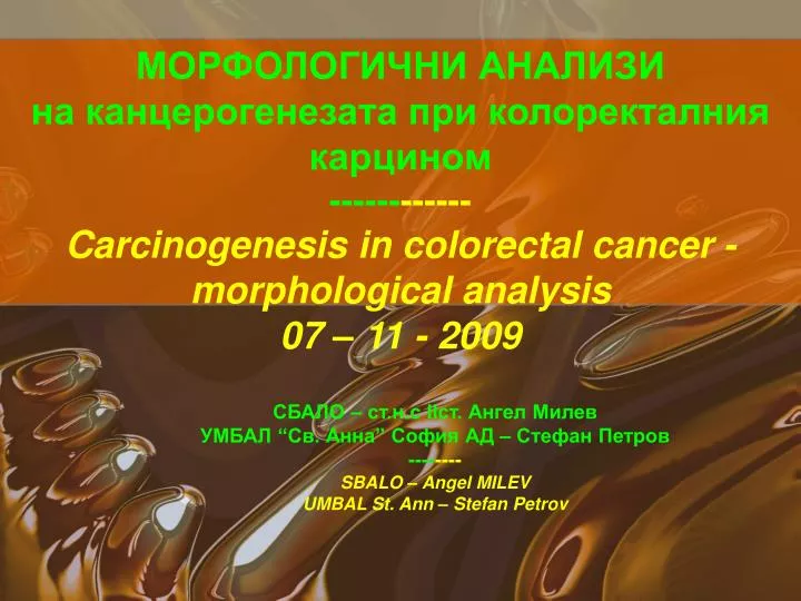
МОРФОЛОГИЧНИ АНАЛИЗИ на канцерогенезата при колоректалния карцином ------ ------ Carcinogenesis in colorectal cancer - morphological analysis 07 – 11 - 2009. СБАЛО – ст.н.с ІІст. Ангел Милев УМБАЛ “Св. Анна” София АД – Стефан Петров ---- ---- SBALO – Angel MILEV UMBAL St. Ann – Stefan Petrov.

E N D
МОРФОЛОГИЧНИ АНАЛИЗИ на канцерогенезата при колоректалния карцином------------Carcinogenesis in colorectal cancer - morphological analysis07 – 11 - 2009 СБАЛО– ст.н.с ІІст. Ангел Милев УМБАЛ “Св. Анна” София АД – Стефан Петров -------- SBALO – Angel MILEV UMBAL St. Ann – Stefan Petrov
Era Uma Vez... (важат и днес) • Основни понятия “CA for clinicians” 1983…
and… • what happens later ?
Kubota & Kino say: 1 2a Аденом с умбиликация в средата (1) Взаимоотношения на честотите на трите вида (3) Аденоматозните жлези формират неравна повърхност (2a) и в част от тях жлезите са атипични (2b) 2b 3 15 %
1 • The American Concept (1999) – C M Fenoglio-Preiser & all.
2 • The American Concept (1999) – C M Fenoglio-Preiser & all.
3 • The American Concept (1999) – C M Fenoglio-Preiser & all. > > > > > > methylene blue ( x 5 ) HE ( x 32 ) Ki 67 ( x 50 ) HE ( x 50 ) > > > >
4 • The American Concept (1999) – C M Fenoglio-Preiser & all.
5 • The American Concept (1999) – C M Fenoglio-Preiser & all. For the first time used the term "serrated" type of polyps Pit-Saw Blade LikePit-Saw Blade Surface
5 • The American Concept (1999) – C M Fenoglio-Preiser & all. ACF & possible relationships between hyperplastic (HP) & adenomatous (AP) polyps
ABC OFCOLORECTAL CANCER (2001) - 1 • Molecular basis for risk factors - Robert G Hardy, Stephen • J Meltzer, Janusz A Jankowski Edited by: D J KERRProfessor & ANNIE M YOUNG, Institute for Cancer Studies, University of Birmingham & F D RICHARD HOBBS >
ABC OFCOLORECTAL CANCER (2001) - 2 • Molecular basis for risk factors - Robert G Hardy, Stephen • J Meltzer, Janusz A Jankowski
Ki-ras; KRAS; Kras – WHO IS IT? • The ras family of oncogenes is constituted of three principal members - K-ras, H-ras and N-ras - all of which have been implicated in the development of human malignancies. The K-ras oncogene resides on chromosome 12p12, and encodes a 21-kD protein (p21ras) involved in the G-protein signal transduction pathway, modulating cellular • proliferation and differentiation.Actually V-Ki-ras2 Kirsten rat sarcoma viral oncogene homolog also known as KRAS is a protein which in humans is encoded by the KRASgene. Like other members of the Ras family, the KRAS protein is a GTPase and is an early player in many signal transduction pathways. • Mutations of the K-ras oncogene result in constitutive activation of this signal transduction pathway and consequently, unregulated proliferation and impaired differentiation. K-ras abnormalities are one of the earliest eventsin the stepwise progression of colorectal neoplasms, being detectable even in histologically unremarkable epithelium and aberrant crypt foci adjacent to cancers. (as shown in the figure > left)
Biomarkers Predicting Clinical Outcome of Epidermal GrowthFactor Receptor - Targeted Therapy in Metastatic ColorectalCancer(S Siena , ASartore-Bianchi , F Di Nicolantonio , JBalfour & A Bardelli) -Milan, Italy 2009 • Overview of interlinked cellular signaling pathways involved in • the proliferation and progression of colorectal cancer. • Agents targeting • signaling proteins that have been evaluated or are currently being evaluated • in phase II, III, or IV clinical trials for colorectal cancer are shown. The • epidermal growth factor receptor (EGFR) – related family of receptor • tyrosine kinases includes human epidermal growth factor receptor • (HER1), EGFR, or c-erbB1; HER2 or c-erbB2; HER3 or c-erbB3; and HER4 • or c-erbB4. C-MET = mesenchymal – epithelial transition factor; EGF = epidermal growth factor; HDAC = histone deacetylases; HGF = hepatocyte growth factor; IGF-1 = insulin-like growth factor-I; IGF-1R = insulin-like growth factor-I receptor; IR = insulin receptor; VEGF = vascular endothelial growth factor; VEGF-R = vascular endothelial growth factor receptor.
KRAS Mutation Testing –Identify metastatic colorectal patients who are non-responsive to EGFR therapy.-1 • The Some of the previous Figure on 3D:
KRAS Mutation Testing –Identify metastatic colorectal patients who are non-responsive to EGFR therapy.-2 • The Basis of the Modern Therapy of CRC - 1
KRAS Mutation Testing –Identify metastatic colorectal patients who are non-responsive to EGFR therapy.-3 • The Basis of the Modern Therapy of CRC - 2
KRAS Mutation Testing –Identify metastatic colorectal patients who are non-responsive to EGFR therapy.-4 • The Visual Image of K-RAS – LUNG ADENO-CA - FLU WHERE IS IT?
KRAS Mutation Testing –Identify metastatic colorectal patients who are non-responsive to EGFR therapy.- 51 • Molecular Pathology Laboratory Network, Inc. (MPLN) offers KRAS mutation testing to assistwith the identification of colorectal cancer (CRC) patients who are most likely to show limited clinical response to anti-epidermal growth factor receptor (anti-EGFR) therapies such as cetuximab (Erbitux®)and panitumumab (Vectibix®). • In January 2009, the American Society of Clinical Oncology released its first Provisional Clinical Opinion recommending that all patients with CRC have tumors tested for KRAS gene mutations prior to the initiation of therapy. • Study Forecasts Savings Using KRAS Test • Based on annual incidence of new metastatic CRC cases,a savings of more than $604 million for cetuximab alone would result through treatment drug stratification based upon KRAS status. • KRAS Mutations Tested • The incidence of KRAS gene mutations in CRC is approximately 35-45%. Identifying patients who will benefit from treatment is vital to ensuring better clinical outcomes as well as determining the best treatment strategies for each patient. • The KRAS mutation test performed at MPLN detects 12 somatic mutations in codons 12 and 13 of the KRAS oncogene. • METHODOLOGY • Tumor tissue is microdissected and DNA extracted from fivemicron sections of formalin-fixed paraffin-embedded tissue • blocks.
KRAS Mutation Testing –Identify metastatic colorectal patients who are non-responsive to EGFR therapy. - 6 • Practical: Test Screen – D-r S. Alexov Private Laboratory • ( sertificated in SWEDEN )
PREDICTING CA RISK IN CR ADENOMA (2009) – 1 (Stavanger, Norway) • Haggitt • classification • Level 0: • Carcinoman situNot • invasive. • Level 1: • CarcinomaInvading • through muscularis • mucosa-limited to the • Head. • Level 2:Carcinoma • neck of the adenoma. • Level 3:Carcinoma • invading anypart of • The stalk. • Level 4:Carcinoma • invading into thethe • submucosa of bowel.
PREDICTINGCA RISK IN CR ADENOMA (2009) – 2(Stavanger, Norway) • Metachronous and synchronous cancer detection. Time from index adenoma detection to cancer detection in synchronous versus metachronous cancer. Synchronous cancer is usually defined as detection at the time of, or within 6 months from, index adenoma detection. Metachronous cancer detection is defined from 6 to 24 months after index detection, but definitions vary considerably in the literature. No universal definitions exist.
PREDICTING CA RISK IN CR ADENOMA (2009) – 3(Stavanger,Norway) • Risk of cancer in adenomas according to type and size. • Derived and modified from WHO
PREDICTINGCA RISK IN CR ADENOMA (2009) – 4(Stavanger, Norway) • Morphology versus morphometry for adenoma assessment. Morphometric measurements performed by quantitative, digitalized assessment of histopathology slides compared with regular pathology assessment through the microscope.
PREDICTINGCA RISK IN CR ADENOMA (2009) – 5(Stavanger, Norway) • Main pathways • in • colorectal • carcinogenesis • represent the • major part • in 75-80% of • patients. • These pathways • have • distinct clinical, • pathological and • genetic • characteristics, • which • can be used for • molecular • classification • for improved • diagnostics, • prognosis and • treatment.
PREDICTINGCA RISK IN CR ADENOMA (2009) – 5(Stavanger, Norway) Bias in biomarker research. Bias may develop at any level, as illustrated in the figure – ranging from clinical, biological or technical standpoints.
OWNSTUDIES – AIM AND M. & M. - 1 - (St.Ann) • AIM: Which of the Polyp isMOST OFTEN SERATED ? • MATHERIAL: • Pinch-biopsy of 241 patients with 404 total lesions, with polyps of the colon, of which 203 single and 201 combined (194 double and 7 triple) were investigated routinely (HE). • METHODS: • In addition to all GIT histochemycal methods, a pilot study of a part of hyperplastic polyps with controls (KRAS) and other was carried out and intestinal adeno-Ca as well – a total of 10 patients. The study was retrospective and the results were processed, using EXCEL 2003 (only %). Graphs were created with the same program.
OWNSTUDIES RESULTS – POLYPS – 1 - (St.Ann) • BASIS OF THE POLYP’S SCIENCE… Hyperplastic Polyps, HE x 32 Tubular Adenoma, HE x 32 Tubulo-Villous Adenoma, HE x 50 Villous Adenoma, HE x 32
OWNSTUDIES RESULTS – POLYPS – 2 - (St.Ann) • BASIS OF THE POLYP’S SCIENCE. Serated Adenomas, HE x 32 Lymphomatoid Polyp, HE x 32 > Melanosis in Peutz-Jeghers Polyp, HE x 32 (arrow) Inflamatory Fibrous Polyp, HE x 32
OWNSTUDIES RESULTS – POLYPS – 3 - (St.Ann) • DYSPLASIAS - LOW & MIDDLE GRADE ( I & II ) > > Dyspl. Gr. I in Hyperplastic Polyp, HE x 32 (arrow) Dyspl. Gr. II in Hyperplastic Polyp, HE x 32 (arrow) > > Dyspl. Gr. II in Serated Polyp, HE x 32 (arrow) Dyspl. Gr. III in Serated Polyp, HE x 32 (arrow)
OWNSTUDIES RESULTS – POLYPS – 4 - (St.Ann) • DYSPLASIAS - HIGH GRADE ( III ) & FOCAL CA., ADENOCA. > > > Dyspl. Gr. III (arrow) &Focal Ca. (2 arrows) in Peutz-Jeghers Polyp, HE x 50 High Differentiation (G1) in INTESTINAL ADENOCA, HE x 32 > Middle Differentiation (G2) in INTESTINAL ADENOCA, HE x 50 (arrow) Low Grade Differentiation (G3) in INTESTINAL ADENOCA, HE x 160
OWNSTUDIES RESULTS – POLYPS – 5 - (St.Ann) • K-Ras TEST – PILOT STUDY (10 patients) – only Hyperplastic Polyps > K-Ras ( - ) in Hyperplastic Polyp, HE x 50 K-Ras ( + ) FOCALLY in Hyperplastic Polyp, HE x 50 (arrow) > > > > K-Ras ( +++ ) FOCALLY in Hyperplastic Polyp, HE x 50 (arrows) K-Ras ( +++ ) FOCALLY in Hyperplastic Polyp, HE x 160 (arrows)
OWNSTUDIES RESULTS – POLYPS – 6 - (St.Ann) • K-Ras TEST – PILOT STUDY (10 patients) – CONTROLS K-Ras ( +++ ) in Focal Ca. - Peutz- Jeghers Polyp, x 160 K-Ras ( +++ ) in G3 INTESTINAL ADENO Ca. , x 160
OWNSTUDIES RESULTS – POLYPS –7 - (St.Ann) (BG) • R E S U L T S – STANDARD METHODS 1. изследвани 38пациенти с общо 115 полипозни лезии ( 63 единични, 52 комбинирани ) * * * *
OWNSTUDIES RESULTS – POLYPS –8 - (St.Ann) (BG) • R E S U L T S – STANDARD METHODS 2. изследвани 203 пациенти с общо 289 полипозни лезии (140 единични, 149комбинирани) * сератни полипи – от общ брой полипи 5,19 % сератни полипи – от общ брой пациенти 7,39 % * * * * * * *
OWNSTUDIES RESULTS – POLYPS –9 - (St.Ann) (BG) R E S U L T S – STANDARD METHODS – COMBINED LESIONS 3. изследвани общо 201 случая с комбинирани полипозни лезии, от които само 7 са тройни * * * * * *
OWNSTUDIES RESULTS – POLYPS –10 - (St.Ann) (BG) • R E S U L T S – K-Ras - PILOT STUDY 4. Ф.кпри-P-J( + ) Туб. Аденом ( +++ ) Адено-Са – G2 ( + ) Хип. П. + Диспл. ІІІ ст. ( + ) Хип. П. + Диспл. ІІІ ст. ( + ++ )
CONCLUSIONS 1. The results clearly indicate a reduction of the percentage of tubular adenomas without dysplasia at the expense of the serated polyps, which corresponds somewhat with the available data of other authors (CM Fenoglio-Preiser & all.) 2. As for the K-Ras test, the only thing that can be assumed about that limited amount of material is, that the rate of the K-Ras (+) cases did not exceed 10%. Part of the surveyed patient samples were with focal expression, unlike other sites (breast, stomach, etc..), which have yet to be specified.
The END • Maybe theKey to the understanding of the colorectal carcinoginesis is here: Maybe the solution to the problem is so close, or not… who knows? But we ought to find it !