Download
1 / 44
440 likes | 460 Views
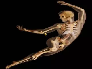
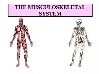
The Musculoskeletal System. Biology for Majors. Types of Skeletons: Hydrostatic. A hydrostatic skeleton is formed by a fluid-filled compartment held under hydrostatic pressure; movement is created by the muscles producing pressure on the fluid. Exoskeleton.

E N D
The Musculoskeletal System Biology for Majors
Types of Skeletons: Hydrostatic A hydrostatic skeleton is formed by a fluid-filled compartment held under hydrostatic pressure; movement is created by the muscles producing pressure on the fluid
Exoskeleton An exoskeleton is a hard external skeleton that protects the outer surface of an organism and enables movement through muscles attached on the inside
Endoskeleton An endoskeleton is an internal skeleton composed of hard, mineralized tissue that also enables movement by attachment to muscles
The Skull The bones of the skull support the structures of the face and protect the brain
Practice Question What is the function of the thoracic cage?
Bone and Calcification • Bone, or osseous tissue, is a connective tissue that constitutes the endoskeleton • It contains specialized cells and a matrix of mineral salts and collagen fibers • The mineral salts primarily include hydroxyapatite, a mineral formed from calcium phosphate • Calcification is the process of deposition of mineral salts on the collagen fiber matrix that crystallizes and hardens the tissue
Long Bones Long bones are longer than they are wide and have a shaft and two ends. The diaphysis, or central shaft, contains bone marrow in a marrow cavity. The rounded ends, the epiphyses, are covered with articular cartilage and are filled with red bone marrow, which produces blood cells
Compact Bone Compact bone tissue is composed of osteons and forms the external layer of all bones
Spongy Bone Spongy bone tissue is composed of trabeculae and forms the inner part of all bones. Trabeculae in spongy bone are arranged such that one side of the bone bears tension and the other withstands compression
Osteoblasts Osteoblasts are bone cells that are responsible for bone formation. Osteoblasts synthesize and secrete the organic part and inorganic part of the extracellular matrix of bone tissue, and collagen fibers. Osteoblasts become trapped in these secretions and differentiate into less active osteocytes
Osteoclasts Osteoclasts are large bone cells with up to 50 nuclei that remove bone structure by releasing lysosomal enzymes and acids that dissolve the bony matrix. These minerals, released from bones into the blood, help regulate calcium concentrations in body fluids. Bone may also be resorbed for remodeling, if the applied stresses have changed
Osteocytes Osteocytes are mature bone cells and are the main cells in bony connective tissue • These cells cannot divide • Osteocytes maintain normal bone structure by recycling the mineral salts in the bony matrix
Osteoprogenitor Cells Osteoprogenitor cells are squamous stem cells that divide to produce daughter cells that differentiate into osteoblasts • Osteoprogenitor cells are important in the repair of fractures
Classification of Joints The functional classification divides joints into three categories: synarthroses, amphiarthroses, and diarthroses: • Synarthroses are a joints that are immovable. This includes sutures, gomphoses, and synchondroses. • Amphiarthroses are joints that allow slight movement, including syndesmoses and symphyses. • Diarthroses are joints that allow for free movement of the joint, as in synovial joints.
Fibrous Joints The bones of fibrous joints are held together by fibrous connective tissue. There is no cavity, or space, present between the bones and so most fibrous joints do not move at all, or are only capable of minor movements. There are three types of fibrous joints: • Sutures • Syndesmoses • Gomphoses
Cartilaginous Joints The bones are connected by cartilage. The two types of cartilaginous joints allow for very little movement: • In a synchondrosis, the bones are joined by hyaline cartilage. Synchondroses are found in the epiphyseal plates of growing bones in children • In symphyses, hyaline cartilage covers the end of the bone but the connection between bones occurs through fibrocartilage. Symphyses are found at the joints between vertebrae
ATP and Muscle Contraction The cross-bridge muscle contraction cycle, which is triggered by Ca2+ binding to the actin active site, is shown. With each contraction cycle, actin moves relative to myosin
Steps of a skeletal muscle contraction: • An action potential reaches the axon of the motor neuron • The action potential activates voltage gated calcium ion channels on the axon, and calcium rushes in • The calcium causes acetylcholine vesicles in the axon to fuse with the membrane, releasing the acetylcholine into the cleft between the axon and the motor end plate of the muscle fiber • The skeletal muscle fiber is excited by large mylenated nerve fibers which attach to the neuromuscular junction. There is one neuromuscular junction for each fiber
Steps of Muscle Contraction Continued • The acetylcholine diffuses across the cleft and binds to nicotinic receptors on the motor end plate, opening channels in the membrane for sodium and potassium. Sodium rushes in, and potassium rushes out. However, because sodium is more permeable, the muscle fiber membrane becomes more positively charged, triggering an action potential • The action potential on the muscle fiber causes the sarcoplasmic reticulum to release calcium ions(Ca++) • The calcium binds to the troponin present on the thin filaments of the myofibrils. The troponin then allosterically modulates the tropomyosin. Normally the tropomyosin physically obstructs binding sites for cross-bridge; once calcium binds to the troponin, the troponin forces the tropomyosin to move out of the way, unblocking the binding sites
More Steps of Muscle Contraction • The cross-bridge (which is already in a ready-state) binds to the newly uncovered binding sites. It then delivers a power stroke • ATP binds the cross-bridge, forcing it to conform in such a way as to break the actin-myosin bond. Another ATP is split to energize the cross bridge again • Steps 7 and 8 repeat as long as calcium is present on thin filament • Throughout this process, the calcium is actively pumped back into the sarcoplasmic reticulum. When no longer present on the thin filament, the tropomyosin changes back to its previous state, so as to block the binding sites again. The cross-bridge then ceases binding to the thin filament, and the contractions cease as well • Muscle contraction remains as long as Ca+2 is abundant in sarcoplasm
Types of Muscle Contractions • Isometric contraction: muscle does not shorten during contraction and does not require the sliding of myofibrils but muscles are stiff • Isotonic contraction: inertia is used to move or work. More energy is used by the muscle and contraction lasts longer than isometric contraction. Isotonic muscle contraction is divided into two categories: • concentric, where the muscle fibers shorten as the muscle contracts (ie. biceps brachialis on the up phase of a biceps curl) • eccentric, where the muscle fibers lengthen as they contract (ie. biceps brachialis on the down phase of a biceps curl) • Twitch: exciting the nerve to a muscle or by passing electrical stimulus through muscle itself. Some fibers contract quickly while others contract slowly • Tonic: maintaining postural tone against the force of gravity
Practice Question Why is it important to eat a diet rich in Calcium?
Quick Review • What are the different types of skeletal systems? • What is the structure and function of bones? • How do Biologists classify the different types of joints on the basis of structure? • What is the role of muscles in locomotion?