Download
1 / 39
400 likes | 558 Views
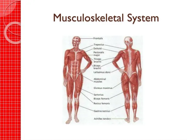
Musculoskeletal System. Common Diagnostic Tests. ANA, antinuclear antibodies Detects SLE, a collagen disease Arthritis can result from SLE Normal = negative Client prep. CRP, C-reactive protein inflammation and auto-immune disorders show abnormal protein

E N D
Common Diagnostic Tests • ANA, antinuclear antibodies • Detects SLE, a collagen disease • Arthritis can result from SLE • Normal = negative • Client prep
CRP, C-reactive protein • inflammation and auto-immune disorders show abnormal protein • Normal = female 1-20, male 1-13mm/h • Can get false negative • Client Prep, usually non-fasting blood draw
Uric Acid-Blood • Elevated with Gout and arthritis • Normal = male 2.1-8.5, female 2.0-6.6 mg/dl • Client prep: usually non-fasting blood draw
Uric Acid-Urine (24 hour collection) • Normal = 250-750 ml/24hr • Client prep
CBC • Hemoglobin • Increase can mean CHF • Decrease can mean SLE or sarcoidosis • Normal = male 14-18, female 12-16 g/dl • Client prep
CBC • WBC • elevated with infection/inflammation • Normal + 5,000 – 10,000/mm3 • Client prep
ESR, Erythrocyte Sedimentation Rate • RBC descent in saline in 1 hour • Increases with inflammation, infection, necrosis, or cancer • Normal = male up to 15, female up to 20 mm/hr • Client prep
RF, Rheumatoid Factor (IgM) • Elevated with autoimmune disease such as Rheumatoid arthritis and SLE • Normal = < 60 U/ml or negative • Client prep
Serum Calcium, detects calcium metabolism • Increase may indicate: metastatic bone tumor, Paget’s disease, acromegaly • Decrease may indicate: rickets, osteomalacia, vitamin D deficiency • Normal = 9.0 – 10.5 mg/dl • < 6mg/dl may lead to tetany (cramps, convulsions, twitching) • > 14mg/dl may lead to coma • Client prep
Radiologic Studies • Arthrogram/Arthrography-Xray with contrast dye into joint to visualize soft tissue of joints (meniscus, ligaments, cartilage)
Arthrogram • Client prep
Arthrogram Procedure: • Cleanse & anesthetize area • Insert needle into joint space • Aspirate fluid to minimize dilution of dye • Leave needle in, replace syringe with dye syringe • Inject contrast and remove needle • ROM to distribute dye • X-rays will be taken • Takes about 30 minutes • May experience some discomfort, pressure, tingling
Following Arthrogram: • Assess for swelling • Apply ice, if needed • Mild analgesic • May hear crepitus after test. This is normal and will disappear in 1-2 days. • Instruct pt to call MD if pain or swelling occurs
CT Scan • X-ray (body scanner) with contrast dye • Three-dimensional cross-sectional view of tissues at various angles • Can identify small differences: Detects edema, hemorrhage, blood flow, infarcts, tumors, infections, aneurysms, demyelinating disease, muscular disease, skeletal abnormalities, disk problems, causes of spinal cord compression • Takes about an hour • Findings as with arthrogram, but 3-D view
CT Scan • Client prep
Following CT Scan • Increase fluid intake to flush dye • Evaluate patient for delayed reaction to dye (usually occurs within 2-6 hours) • Treat with antihistamine and/or steroids, if indicated
CT Scan Procedure • Patient must lie still • Show picture of CT machine and discuss claustrophobia, may need antianxiety med • Performed by a radiologist • Takes 30-45 minutes • Discomfort includes lying still on a hard surface, peripheral venipuncture, mild nausea, salty taste, flushing, and warmth from dye
MRI • MRI/Magnetic Resonance Imaging • Magnetic field and radio waves, noninvasive • Can evaluate soft & hard tissue, & blood vessels • Unique d/t no exposure to ionizing radiation • Advantages over CT • Disadvantages
MRI • Contraindications: • > 300 lbs • Claustrophobia • Metal implants, clips, pacemaker, infusion pumps • Pregnancy (long-term effects not known) • If on continuous life support
Client prep: • Obtain consent • Can drive afterwards without assistance • Assess for contraindications • Show picture of machine, discuss claustrophobia • Remove all metal objects from body (create artifacts, can go flying, damages credit cards) • Must remain motionless in supine position • Will hear thumping sound, ear plugs available • Empty bladder prior to test for comfort • No food or fluid restrictions prior to test • Explain procedure
MRI Procedure • Lie flat on hard table that slide into a tube • Must lie still • Can talk to or listen to staff • Magnevist (contrast agent) may be used via IV • Performed by radiologist • Takes 30 to 90 minutes • Discomfort from lying on hard surface, possible venipuncture, possible tingling in teeth (metal fillings) • No postprocedural care needed
MRI • Detects: edema, hemorrhage, blood flow, infarcts, tumors, infections, aneurysms, demyelinating disease, muscular disease, skeletal abnormalities, disk problems, causes of spinal cord compression
X-ray • X-ray, electromagnetic radiation passes photons (light particles) through the body onto film • Bone (very dense) blocks photons, appears white • Air appears black • Muscle, fat, and fluid appear as various shades of gray • Metal and contrast block almost all photons and appear bright white
X-ray • Client prep • Nonfasting • Position determined by area to be x-rayed • Patient should be still, usually hold breath • Contraindicated if pregnant • May need to remove jewelry & don a gown • No discomfort except r/t position • Detects fractures and some joint abnormalities
Myelogram • Myelogram • X-ray with contrast dye of spinal subarachnoid space • Detects spinal tumors, herniated discs, bone spurs, cervical ankylosing spondylosis, arthritic lumbar stenosis • Contraindications: Multiple sclerosis patients (may cause exacerbation), ICP, infection near lumbar puncture sight, allergy to shellfish
Myelogram • Client prep
Myelogram Procedure • Empty bladder • A lumbar puncture is performed • 15 ml CSF removed • 15 ml of radiopaque dye injected • Patient will be tilted up and down to spread dye (prone position) • Lights are off, dye followed with fluoroscopy • X-ray films taken • Needle remains in place until exam concluded • Done by radiologist and takes 45 minutes • Discomfort varies from mild to severe
Following Myelogram • Bed rest for several hours • Head position varies per dye used, per MD order • Monitor for bleeding, fever, headache, photophobia, seizure, VS, ability to void, reaction to dye • Possible med restrictions • Push fluids
Bone Scan • Bone Scan • Radioactive isotope intravenous • They use a gamma camera to detect “hot spots” of activity where the isotope collects • Can detect tumor, arthritis, fracture, necrosis, degenerative changes, osteomyelitis • Normal = uniform distribution • Abnormal = area of higher concentration • Contraindicated in pregnancy, breastfeeding
Bone Scan • Advantages: • Disadvantages:
Bone Scan • Client prep: • Explain procedure, is non-fasting, no sedation required • Arrive at Nuclear Medicine department 4 hours prior to test • Dye given IV, takes 4 hours to travel to bones • Push fluid to aid in dye distribution • Empty bladder upon return to avoid artifact • You may be asked to wear a gown • Done in supine, prone, & lateral position, takes an hour • Takes 6-24 hours for dye to leave system (push fluids) • Discomfort is needle stick for dye infusion, and hard surface
Bone Mineral Density/BMD • BMD • Measures bone mass • The only test to diagnose osteoporosis • Normal is comparative to same age, sex, size. Lower density = higher risk for fractures • -1 to –2, Osteopenia • < -2.5, Osteoporosis • Client prep: Non-fasting, non-invasive, do Q2 yrs
Other Tests • Arthrocentesis • Obtain synovial fluid from a joint • Needle aspiration • Sterile procedure • Detects infections, synovitis, crystal-induced arthritis, tumors, joint degeneration • Inject anti-inflammatory medications • Normal= Clear, straw-colored fluid, no crystals • Contraindicated if infection near joint being tested
Arthrocentesis cont’d • Informed consent • Explain procedure • May or may not be fasting • Local anesthetic • Aseptic procedure • Fluid may be removed, Steroid may be injected • Apply pressure dressing following procedure • May do venipuncture to compare chemical content • Doctor office or bedside, by MD, takes 10 minutes • Pain may worsen after test
Following Arthrocentesis • Assess for pain, fever, swelling • Apply ice • Apply pressure dressing to decrease reaccumulation of fluid or hematoma • Avoid strenuous use of joint for several days
Arthroscopy-used most often for knee • Small incision, endoscope • Examine the inside of a joint • Diagnose disease, meniscus problems, torn cartilage, remove small bodies, do biopsy • Advantage: allows direct visualization, can perform surgery, can monitor disease progress, can attach video camera; can examine, biopsy, or do surgery • Contraindications:Infection or ankylosis in joint
Arthroscopy • Client prep: • Obtain consent • NPO at midnight • Teach crutch use for post procedure use • Shave 6” above and below joint • May use local or general anesthesia • Pressure wrap or tourniquet • Knee at 45 degree angle • May have 2-3 small incision sights • Sutured with dressing applied • Done by orthopedic surgeon, takes 15 to 30 minutes
Arthroscopy Follow-up • Asses neurologic status and circulatory status • Assess for sxs of infection, for drainage • Teach to elevate & ice to decrease swelling • May walk with crutches if MD order • Suture removal in 7-10 days