Download
1 / 47
470 likes | 671 Views
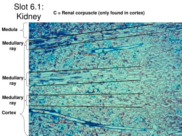
Slot 6.1: Kidney. C = Renal corpuscle (only found in cortex). Medula. Medullary ray. Medullary ray. Medullary ray. Cortex. Slot 6.2: Diagram of nephron blood supply. G = glomerulus. Vasa recta. V = vascular pole. Slot 6.3: Renal Corpuscle. C = Bowman’s capsule at urinary pole.

E N D
Slot 6.1: Kidney C = Renal corpuscle (only found in cortex) Medula Medullary ray Medullary ray Medullary ray Cortex
Slot 6.2: Diagram of nephron blood supply G = glomerulus Vasa recta
V = vascular pole Slot 6.3: Renal Corpuscle C = Bowman’s capsule at urinary pole A= afferent arteriole Arrowhead = macula densa Juxtaglomerular cells Distal tubule D = distal tubule P=proximal tubule Dark cells = podocytes
Slot 6.4: Juxtaglomerular apparatus Arrowheads = juxtaglomerular cells a = afferent arteriole (vascular pole) e = endothelial cell p = podocyte (right) p = Proximal tubule (left) m = macula densa Renal corpuscle d=distal tubule U = Urinary pole
Slot 6.5: Kidney a = vasa recta c = collecting duct d = distal tubules p = proximal tubules Arrows = Brush border Arrowheads = intercalated cells
a = vasa recta Slot 6.6: Kidney (medulla) v = venule (vasa recta) i = interstitial CT C = collecting duct H = thick loop of Henle T = thin loop of henle
Slot 6.7: Ureter Arrows = binucleate cells (diagnostic of transitional epithelium) Adventitia Transitional epithelium Mucosa CT Muscularis
Slot 6.8: Bladder Transitional epithelium Lamina propria
Slot 6.9: Diagram of Ovary Secondary follicles Graffian follicle Primary follicle Primordial follicles Corpus luteum
Slot 6.10: Ovary sections P = primordial follicles G = primary follicles Arrows = zona pellucida (left) Arrows = theca folliculi (right) Granulosa cells Granulosa cells
Slot 6.11: Fallopian tube Arrows = cilia on epithelial cells moves egg through oviduct LP = lamina propria
Slot 6.13: Uterine endometrium sections Proliferative Phase Secretory Phase Compact Layer Myometrium Endometrium Spongy layer
Slot 6.14: Uterine endometrium sections Early menstruation (right) - surface layers have been shed Progestational stage (left) Spongy layer Basal layer B = basal layer
Maternal component Slot 6.16: Diagram of placental villi Fetal component
Slot 6.17: Placenta sections Arrows = fetal capillaries (vascularization) Eigth Month (left) Ninth Month (right) Hofbauer cells Syncitial trophoblast
Slot 6.18: Uterine Cervix Simple columnar Stratied squamous C=cervix Vagina Plicate palmatae
Pre-puberty Pregnancy gland Slot 6.19: Mammary gland Duct Later pregnancy During Lactation Glands: colostrum Milk
Slot 6.20: Kidney Cortex Medulla Renal corpuscles Medullary rays
Slot 6.22: Kidney cortex Distal convoluted tubules Macula densa R = Renal corpuscles Afferent arteriole R R Proximal convoluted tubule Urinary pole
Slot 6.23: Kidney cortex Renal corpuscles Medulary ray
Slot 6.24: Kidney medulla Collecting duct Thick Loop of Henle Thin loops of Henle
Slot 6.25: Kidney medulla Thin loop of Henle Collecting duct Thick loop of Henle
Slot 6.26: Renal corpuscle Vascular pole Urinary pole Proximal tubule Macula densa
Slot 6.27: Renal corpuscles Shows vascularization of the glomeruli
Slot 6.28: Renal corpuscle Macula densa Vascular pole Urinary pole
Slot 6.29: Renal corpuscles RC RC Collecting duct (pale cells) Proximal tubule Distal tubule
Slot 6.30:Kidney Medulla Simple cuboidal cells of collecting duct
Slot 6.33: Kidney medulla Thick loop of Henle Collecting ducts Thin loop of Henle
Slot 6.34: Fetal kidney (largely inactive since fetus relies on maternal filtration) Medulla Cortex Glomeruli
Slot 6.35: Ovary Germinal epithelium Medulla Mesovarium Cortex Graffian (mature) follicle Corpus Luteum
Slot 6.36: Ovarian cortex Zona pellucida Primary follicle Theca folliculi Antrum Secondary follicle Granulosa cells Early secondary
Slot 6.38: Ovarian follicle Zona pellucida Primary follicle Theca folliculi Corpus Luteum
Lamina propria Slot 6.39:Fallopian tube (ciliated) PSC w/ cilia PSC w/ cilia
Slot 6.40: Uterine endometrium, Secretory Phase Glands - secrete carbohydrate rich fluid to nourish embryo
Slot 6.41: Uterine endometrium, menstrual stage Note rough edges from sloughing off Basal layer Myometrium
Slot 6.42: Placental villi Syncitial trophoblast layer Areolar CT with capillaries
Slot 6.43: Cervix Stratified squamous Plicae palmatae
Slot 6.44: Mammary Gland Alveoli Connective Tissue (CT) Lobule
Slot 6.45: Nipple and areolar gland Sinus Lactiferous duct Areolar Gland
Slot 6.46: Nipple, cat Lactiferous sinus Nipple Alveolar glands
Slot 6.47: Mammary gland (early pregnancy) Glands CT CT