Download
1 / 64
670 likes | 989 Views
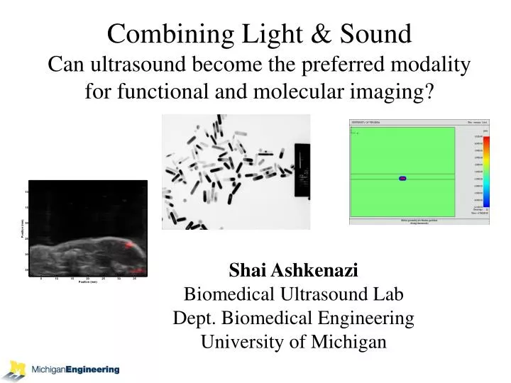
Combining Light & Sound Can ultrasound become the preferred modality for functional and molecular imaging?. Shai Ashkenazi Biomedical Ultrasound Lab Dept. Biomedical Engineering University of Michigan. Imaging devices. Imaging methods. Photoacoustic. Ultrasound. Imaging agents. Outline.

E N D
Combining Light & SoundCan ultrasound become the preferred modality for functional and molecular imaging? Shai Ashkenazi Biomedical Ultrasound Lab Dept. Biomedical Engineering University of Michigan
Imaging devices Imaging methods Photoacoustic Ultrasound Imaging agents Outline
Ultrasound Imaging • Array of Tx/Rx elements • Beam steering and focusing – time delayed channel excitation • Receive – delay & sum • Reflections – different density, speed of sound
Ultrasound Imaging 100 Penetration depth (mm) Low MHz 10 – 20 MHz 10 > 20 MHz (UBM) Abnormal Thyroid Gland 1 0.01 0.1 1 Resolution (mm)
Optoacoustic US Transducers Receive / Transmit Hi Q
Bell’s Photophone February 1880
Etalon detector – principle of operation Etalon PD Array (camera) CW laser
Ultrasound – Space/time load PD Array (camera) 2Kg CW laser Etalon detector – principle of operation Etalon
Piezo vs. EtalonComparison of sensitivity Pulse-Echo Etalon Amplitude 0 0 2.7 2.9 5.3 5.5 5.7 Time (s) Time (s) PIEZO TRANS ETALON
Optical Generation of Ultrasound Water Black PDMS Clear PDMS Laser pulse • High thermal expansion • Optically absorbing
2D Gold Nanostructure 4.5 um PDMS layer 128 nm 20 nm 220 nm Glass Substrate
Acoustic pressure increases linearly with optical input energy Thermal damage threshold: 25 uJ delivered to a spot size of 25 um Acoustic pressure at thermal damage threshold: 500 kPa at 10 mm Acoustic Pressure
Integrated Device SU-8 protection layer PDMS layer Etalon 6 um 200 nm Ultrasound Generation Beam Ultrasound Detection Beam
Output = T + S l Resonance optics T S T = - S (critical coupling) S = 0 (off-resonance phase cancelation)
Ultrasound Transducer US Pulser Transmission c a b (nm) 1558 1563 0 16.5 18 Time (s) Experimental verification Tunable Laser Photodetector
0 Trans. Modulation Transmission c 0 a b (nm) 1558 1563 0 16.5 18 Time (s) Wavelength dependence 10 MHz Transducer
Fiber coupled optical circulator λm λ1 λ2 λ3 … In Tunable laser Out λm+1 … λ2m Demux and Photodetector array Array configurations 80 elements sharing 1 waveguide 2D Arrays Demultiplexer and Photodetector array Miniaturization of high-Freq arrays for intravascular and “in-vivo” microscope application
Micron size elements High frequency arrays > 30 MHz High SNR (size independent noise) Wide Bandwidth > 50 MHz Selectable sensitivity “Shiftable” dynamic range High BW signal comm. (80 Ch. on SMF using 100 GHz standard grid) Why Micro-Optics for Ultrasound Devices?
High resolution ultrasound microscopy at the tip of a needle Guiding biopsy Reducing bleeding complications (e.g. in kidney biopsy) Applications – Smart Needle 200 µm Receive r array Transmitter 2 mm Side viewing G23 0.64 mm
500 mm Photoacoustic Imaging
PA imaging • Laser pulse (~5 ns) • Heat absorption • Temp. rise (~ 0.01 °C) • Thermal expansion (strain ~ 10-5) • Acoustic propogation • Detection and Source reconstruction Receiver
Etalon PD Array (camera) CW laser Etalon for Photoacoustic imaging
2D phantom imaging 100 mm Photoacoustic image 0.11mm Optical image
500 mm Nerve cord imaging Nerve Cord In Lobster Tail 532 nm pulsed illumination Probe laser scan lines (4mm x 0.36mm aperture)
3D phantom imaging 50 µm Array size: 128x128 Element spacing: 30 um
Pig Coronary Artery 700 nm Axial Position (mm) Lateral Position (mm)
Bioconjugation Gold Nanorod Surfactant (CTAB) Antibody PAA
Cell Culture Setup AM OS UT SC CC BX Laser OPO
Photoacoustic Image – LNCaP Cells 0 -10 Conjugated Nanorods -20 -30 1 mm -40 0 -10 Unconjugated Nanorods -20 -30 -40
UltraSound-PhotoAcoustic (USPA) ImagingCombined Modality US UA PH BX Laser OPO SYNC
Photoacoustics provides an exciting vehicle for molecular imaging PEBBLES can be detected at only 10 particles per cell with 100 nm particle diameter Nanorods can be detected at only 50 particles per cell with volume 50 times less than PEBBLE Both agents can be made much more efficient Conclusions
Optical resonators for ultrasound sensing PA contrast for cancer detection Sensor dyes for functional PAI PA sensor for protease activity Future research projects
Ultimate sensitivity for PAI applications – Acoustic noise limited Explore structures for optimal acousto-optic interaction Membrane interface Air-water interface Optical resonators for ultrasound sensing Waveguide Bragg Grating
Real-time PA imager Small animals Clinical trials Stability-dynamics of nanoparticles in-vivo Cell targeting PA contrast for cancer detection - Prostate Cancer - Thyroid cancer
Develop PA imaging of pH, Ca, O2, and other Study PA sensing mechanisms Absorption (change, spectral shift) Fluorescence quenching PA increase Life time of non-radiative decay change in PA shape Delivery agents - Dye embedded nanoparticles Sensor dyes for functional PAI Combine versatility of molecular probes with PAI
Example - pH dye SNAFR-5F
T H AN K S EECS Jay Guo ChungYen Chao Tao ling JingSung Chemistry Raoul Kopelman Gwangseong Kim Tom Horvath Rodney Agayan Chemical Eng. Nick Kotov Ashish Agarwal Cancer Center Mark Day Kathleen Day