Download
1 / 49
520 likes | 793 Views
Bone. Prepared by Dr.Salah Mohammad Fateh MBChB,DMRD,FIBMS(radiology). Lecture no. 1. Vertebral column. Lecture no. 1. Radiological approach to Bone diseases. Prepared by Dr.Salah Mhamad Fateh MBChB,DMRD,FIBMS(radiology). Imaging techniques;. X-ray Isotope US CT MRI.

E N D
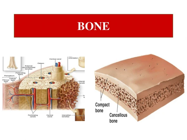
Bone Prepared by Dr.Salah Mohammad Fateh MBChB,DMRD,FIBMS(radiology) Lecture no. 1
Lecture no. 1 Radiological approach to Bone diseases Prepared by Dr.Salah Mhamad Fateh MBChB,DMRD,FIBMS(radiology)
Imaging techniques; • X-ray • Isotope • US • CT • MRI
Initially, a musculoskeletal lesion should be simply imaged with a plain film. It should be remembered that plain films remain the most reliable imaging method for assessment of both biological activity and probable histological diagnosis of an osseous lesion.
1-Decreased bone density 2-Increased bone density (sclerosis). 3- Periosteal reaction 4- Cortical thickening 5- Alteration in the trabecular pattern 6- Alteration in the shape of a bone 7-Altreration in bone age
1-Decreased bone density • Localize (lytic area or area of ‘ bone distruction’) • Generalize
2-increased bone density (sclerosis). • Focal • Generalized
3- periostial reaction; • It refers to excessive bone produced by the periosteum, which occur in response to infection , trauma & tumors
4- Cortical thickening • Also involve laying down of new bone by the periosteum,but the process is very slow & it has the same homogeneous density as does the normal cortex& there is no separate lines or specules of calcification as seen in a periosteal reaction
causes 1- chronic osteomyelitis. 2-healed trauma 3- response to chronic stress or benign tumor
5- alteration in the trabecular pattern Usually involving a reduction in the no. of trabeculae with an alteration in the remaining trabeculae. e.g in osteoporosis, there is reduction in the no. of the trabeculae & remaining trabiculae are more prominent than usual associated with thinning of the cortex. in paget‘s disease , there is thickening of the trabeculae & associated with thickening of the cortex & bone expansion
Normal Local osteoporosis ( femur)
6- alteration in the shape of a bone • Congenital • Acquired , e.g Acromegaly, expanding bone tumors
2-US in musculoskeletal disease US can not demonstrate bone pathology but does have a complementary imaging role; • Detecting tenosynovitis, tendon tear & rupture. • In diagnosis of arthritis & osteomyelitis
3-Radionuclide bone scaning Technetium-99m lablled phosphate complexes
Indication of radionuclide scan are • Detection of metastasis. • Detection of osteomyelitis . • Determination of whether a lesion in solitary or multiple. • Investigation of clinically suspected lesion when the Plain radiograph is –ve. • Investigation of radiographically equivocal cases whether is significant or not. • Investigation of pain full prosthesis .
4-CT in bone disease • Is only needed in selected cases. • Indications for bone CT are • Demonstrating abnormalities in the areas where interpretation of plain films are frequently difficult for exam. Spine , hip &pelvis • As a guide for bone biopsy. • Demonstration of the extent &characterization of the bone tumor in selected cases to complement MRI
4-MRI(magnetic resonance imaging) • Play a vital important role in musculoskeletal disorders. • In can demonstrate bone marrow directly but calcified tissues & cortical bones produces no signal. • MRI particularly good for showing soft tissue abnormalities
Indications of MRI • Disc herniation & spinal cord or nerve roots compression. • Dx of bone metastasis. • Extend of primary bone tumor. • To image soft tissue masses • To Dx osteomyelitis & shows any soft tissue abscess. • To Dx avascular necrosis & other joint pathologies