Download
1 / 27
270 likes | 289 Views
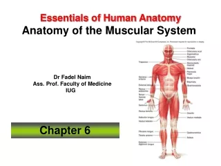
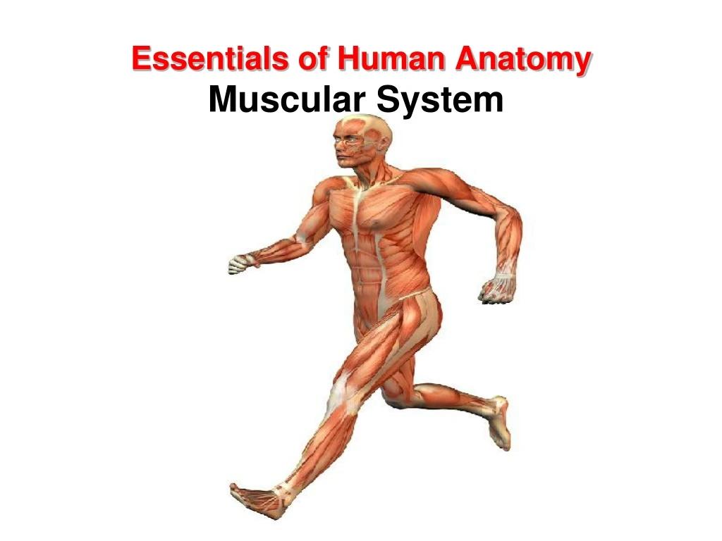
Essentials of Human Anatomy Muscular System. Introduction. There are more than 600 skeletal muscles in the body From 40% to 50% of body weight is skeletal muscle Muscles, along with the skeleton, determine the form and contour of the body. Functions of skeletal Muscles. Body movements

E N D
Introduction • There are more than 600 skeletal muscles in the body • From 40% to 50% of body weight is skeletal muscle • Muscles, along with the skeleton, determine the form and contour of the body
Functions of skeletal Muscles • Body movements • Maintaining Posture • Stabilizing Joints • Body positions • Generating heat
Muscular System • Three basic muscle types are found in the body • Skeletal muscle • Cardiac muscle • Smooth muscle
4 Unique Characteristics of Muscle Tissue • Excitability is equated with responsiveness. • Contractility causes the fiber to shorten resulting in either a pull on bones or the movement of specific body parts. • Elasticity is the muscle’s ability to return to its original length when tension is released. • Extensibility is capability of extending in length in response to the contraction of opposing muscle fibers.
Structure of a Skeletal Muscle • Skeletal Muscle • Organ of the muscular system • - skeletal muscle tissue • - nervous tissue • - blood • - connective tissues • Fascia • Tendons • Aponeuroses
Skeletal Muscle Tissue • Skeletal muscles are organs • Vary in shape and size • A skeletal muscle is composed of cells • Each cell is as long as the muscle • Small muscle: • 100 micrometers long; 10 micrometers in diameter • Large muscle: • 35 centimeters long; 100 micrometers in diameter
Skeletal Muscle Structure • Connective tissue components • Endomysium Delicate connective tissue membrane that covers specialized skeletal muscle fibers • Perimysium Tough connective tissue binding together fascicles • Epimysium Coarse sheath covering the muscle as a whole • These three fibrous components may become a tendon or an aponeurosis
Muscle Fiber Structure • Multiple nuclei • Sarcolemma • T-tubules • Sarcoplasmic reticulum • Sarcoplasm • Mitochondria • Glycogen & ions • Myofibrils
Neuromuscular Junction • Also known as myoneural junction • Site where an axon and muscle fiber meet • Motor neuron • Motor end plate • Synapse • Synaptic cleft • Synaptic vesicles • Neurotransmitters
Motor Unit • single motor neuron • all muscle fibers controlled by motor neuron
Types of Contractions • Eccentric – lengthening contraction • Concentric – shortening contraction • Isometric – muscle contracts but does not change length • Isotonic – muscle contracts and changes length
Muscle Atrophy • Reduction in muscle size, tone, and power. • Due to reduced stimulation, it loses both mass and tone. • Muscle becomes flaccid, and its fibers decrease in size and become weaker. • Even a temporary reduction in muscle use can lead to muscular atrophy.
Muscle Hypertrophy • An increase in muscle fiber size. • Muscle size may be improved by exercising. • Repetitive, exhaustive stimulation of muscle fibers results in more mitochondria, larger glycogen reserves, and an increased ability to produce ATP. • Ultimately, each muscle fiber develops more myofibrils, and each myofibril contains a larger number of myofilaments.
Posture • Maintaining the posture of the body is one of the major roles muscles play • “Good posture”—body alignment that most favors function and requires the least muscular work to maintain, keeping the body’s center of gravity over its base
Posture • How posture is maintained • Muscles exert a continual pull on bones in the opposite direction from gravity • Structures and systems other than muscle and bones have a role in maintaining posture • Nervous system—responsible for determining muscle tone and also regulation and coordination of the amount of pull exerted by individual muscles • Respiratory, digestive, excretory, and endocrine systems all contribute to maintain posture
Axial Muscles • Have both their origins and insertions on parts of the axial skeleton. • Support and move the head and spinal column. • Function in nonverbal communication by affecting facial features. • Move the lower jaw during chewing. • Assist in food processing and swallowing. • Aid breathing. • Support and protect the abdominal and pelvic organs. • Are not responsible for stabilizing or moving the pectoral or pelvic girdles or their attached limbs.
Appendicular Muscles • Control the movements of the upper and lower limbs. • Stabilize and control the movements of the pectoral and pelvic girdles. • Organized into groups based on their location in the body or the part of the skeleton they move. • Work in groups that are either synergistic or antagonistic.
Appendicular Muscles • Organized into specific groups. • muscles that move the pectoral girdle • muscles that move the glenohumeral joint/arm • arm and forearm muscles that move the elbow joint/forearm • forearm muscles that move the wrist joint, hand, and fingers • intrinsic muscles of the hand
Intramuscular Injections • The gluteus maximus is a large, thick muscle with coarse Fasciculi that can be easily separated without damage. • The great thickness of this muscle makes it ideal for intramuscular Injections. • To avoid injury to the underlying Sciatic nerve, the injection should be given well forward On the upper outer quadrant of the buttock.