Download
1 / 46
470 likes | 603 Views
Essentials of Human Anatomy. Respiratory System. 1. Organization and Functions of the Respiratory System. Structural classifications: upper respiratory tract lower respiratory tract . Functional classifications: Conducting portion: transports air. Nose nasal cavity Pharynx Larynx

E N D
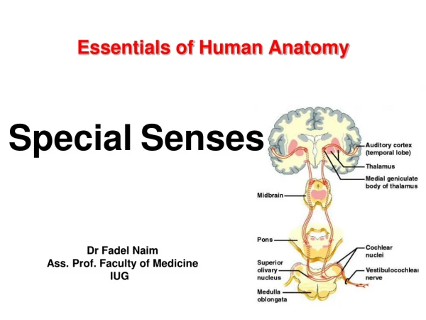
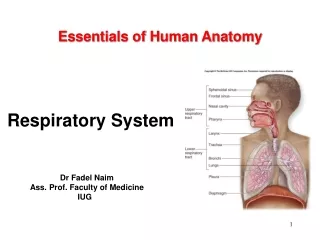
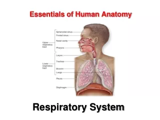
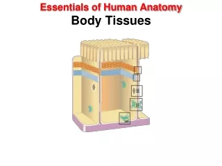
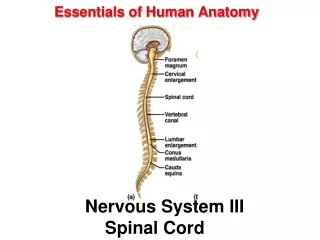
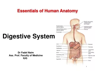
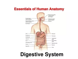
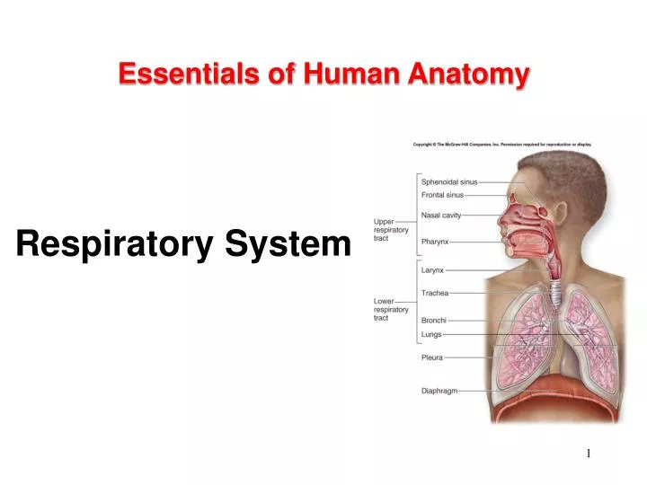
Essentials of Human Anatomy Respiratory System 1
Organization and Functions of the Respiratory System Structural classifications: • upper respiratory tract • lower respiratory tract. • Functional classifications: • Conducting portion: transports air. • Nose • nasal cavity • Pharynx • Larynx • Trachea • progressively smaller airways, from the primary bronchi to the bronchioles
Organization and Functions of the Respiratory System • Functional classifications: • Respiratory portion: carries out gas exchange. • respiratory bronchioles • alveolar ducts • air sacs called alveoli • Upper respiratory tract is all conducting • Lower respiratory tract has both conducting and respiratory portions
Respiratory System Functions • Breathing (pulmonary ventilation): • consists of two cyclic phases: • inhalation, also called inspiration • exhalation, also called expiration • Inhalation draws gases into the lungs. • Exhalation forces gases out of the lungs. • Gas exchange: O2 and CO2 • External respiration • External environment and blood • Internal respiration • Blood and cells
Respiratory System Functions • Gas conditioning: • Warmed • Humidified • Cleaned of particulates • Sound production: • Movement of air over true vocal cords • Also involves nose, paranasal sinuses, teeth, lips and tongue • Olfaction: • Olfactory epithelium over superior nasal conchae • Defense: • Course hairs, mucus, lymphoid tissue
Upper Respiratory Tract • Composed of • the nose • the nasal cavity • the paranasal sinuses • the pharynx (throat) • and associated structures. • All part of the conducting portion of the respiratory system.
Paranasal Sinuses • Paranasal sinuses: • In four skull bones • paired air spaces • decrease skull bone weight • Named for the bones in which they are housed. • frontal • ethmoidal • sphenoidal • maxillary • Communicate with the nasal cavity by ducts. • Covered with the same pseudostratified ciliated columnar epithelium as the nasal cavity.
Pharynx • Common to both the respiratory and digestive systems. • Commonly called the throat. • Funnel-shaped • slightly wider superiorly and narrower inferiorly. • Originates posterior to the nasal and oral cavities • Extends inferiorly near the level of the bifurcation of the larynx and esophagus. • Common pathway for both air and food.
Pharynx • Walls: • lined by a mucosa • contain skeletal muscles primarily used for swallowing. • Flexible lateral walls • distensible • to force swallowed food into the esophagus. • Partitioned into three adjoining regions: • nasopharynx • oropharynx • laryngopharynx
Lower Respiratory Tract • Conducting portion • Larynx • Trachea • Bronchi • bronchioles and their associated structures • Respiratory portion of the respiratory system • respiratory bronchioles • alveolar ducts • alveoli
Larynx • Short, somewhat cylindrical airway • Location: • bounded posteriorly by the laryngopharynx, • inferiorly by the trachea. • Prevents swallowed materials from entering the lower respiratory tract. • Conducts air into the lower respiratory tract. • Produces sounds.
Sound Production • Two pairs of ligaments • Inferior ligaments, called vocal ligaments • covered by a mucous membrane • vocal folds: ligament and mucosa. • are “true vocal cords” • they produce sound when air passes between them • Superior ligaments, called vestibular ligaments • Covered by mucosa • vestibular folds: ligament and mucosa • Are “false vocal cords” • no function in sound production • protect the vocal folds. • The vestibular folds attach to the corniculate cartilages.
Sound Production • The tension, length, and position of the vocal folds determine the quality of the sound. • Longer vocal folds produce lower sounds • Loudness based on force of air • Rima glottidis: opening between the vocal folds • Glottis: rima glottidis and the vocal folds
Tracheostomy Performed to allow air to bypass an obstruction within the larynx
Trachea • A flexible, slightly rigid tubular organ • often referred to as the “windpipe.” • Extends through the mediastinum • immediately anterior to the esophagus • inferior to the larynx • superior to the primary bronchi of the lungs. • Anterior and lateral walls of the trachea are supported by 15 to 20 C-shaped tracheal cartilages. • cartilage rings reinforce and provide some rigidity to the tracheal wall to ensure that the trachea remains open (patent) at all times • cartilage rings are connected by elastic sheets called anular ligaments
Trachea • At the level of the sternal angle, the trachea bifurcates into two smaller tubes, called the right and left primary bronchi. • Each primary bronchus projects laterally toward each lung. • The most inferior tracheal cartilage separates the primary bronchi at their origin and forms an internal ridge called the carina.
Tracheal Blockage Heimlich Maneuver or abdominal thrust
Bronchial Tree • A highly branched system • air-conducting passages • originate from the left and right primary bronchi. • Progressively branch into narrower tubes as they diverge throughout the lungs before terminating in terminal bronchioles. • Primary bronchi • Incomplete rings of hyaline cartilage ensure that they remain open. • Right primary bronchus • shorter, wider, and more vertically oriented than the left primary bronchus. • Foreign particles are more likely to lodge in the right primary bronchus.
Bronchial Tree • Primary bronchi • enter the hilum of each lung • Also entering hilum: • pulmonary vessels • lymphatic vessels • nerves. • Secondary bronchi (or lobar bronchi) • Branch of primary bronchus • left lung: • two lobes • two secondary bronchi • right lung • three lobes • three secondary bronchi. • Tertiarybronchi (or segmental bronchi) • Branch of secondary bronchi • left lung is supplied by 8 to 10 tertiary bronchi. • right lung is supplied by 10 tertiary bronchi • supply a part of the lung called a bronchopulmonary segment.
From Bronchi to Lungs • 1 bronchi (enter lungs at hilus, complete cartilage rings) • 2 bronchi (from now on cartilage plates) • 3 bronchi • Bronchioles • Terminal bronchioles • Respiratory bronchioles • Alveolar ducts • Alveolar sacs Conducting portion Respiratory portion
Alveolar Organization Alveoli are site of gas exchange Close association with capillaries Lots of elastic fibers in alveolar wall Alveolar cells Type I cells – respiratory epitheliocytes Type II cells – septal cells – produce surfactant Alveolar Macrophages – dust cells – phagocytic
Respiratory Bronchioles, Alveolar Ducts, and Alveoli • Contain small saccular outpocketings called alveoli. • An alveolus is about 0.25 to 0.5 millimeter in diameter. • Its thin wall is specialized to promote diffusion of gases between the alveolus and the blood in the pulmonary capillaries. • Gas exchange can take place in the respiratory bronchioles and alveolar ducts as well as in the lungs, which contain approximately 300–400 million alveoli. • The spongy nature of the lung is due to the packing of millions of alveoli together.
Gross Anatomy of the Lungs • Each lung has a conical shape. • Its wide, concave base rests upon the muscular diaphragm. • Its relatively blunt superior region, called the apex or (cupola), projects superiorly to a point that is slightly superior and posterior to the clavicle. • Both lungs are bordered by the thoracic wall anteriorly, laterally, and posteriorly, and supported by the rib cage. • Toward the midline, the lungs are separated from each other by the mediastinum. • The relatively broad, rounded surface in contact with the thoracic wall is called the costal surface of the lung.
Pleura and Pleural Cavities • The outer surface of each lung and the adjacent internal thoracic wall are lined by a serous membrane called pleura, which is formed from simple squamous epithelium. • The outer surface of each lung is tightly covered by the visceral pleura, while the internal thoracic walls, the lateral surfaces of the mediastinum, and the superior surface of the diaphragm are lined by the parietal pleura. • The parietal and visceral pleural layers are continuous at the hilum of each lung.
Pleura and Pleural Cavities • The outer surface of each lung is tightly covered by the visceral pleura, while the internal thoracic walls, the lateral surfaces of the mediastinum, and the superior surface of the diaphragm are lined by the parietal pleura. • The potential space between these serous membrane layers is a pleural cavity. • The pleural membranes produce a thin, serous fluid that circulates in the pleural cavity and acts as a lubricant, ensuring minimal friction during breathing.
Respiratory Muscles Diaphragm: depresses inhalation External intercostals: elevate ribs inhalation Internal intercostals: depress ribs active exhalation Accessory muscles - serratus anterior, scalenes, pectoralis minor, sternocleidomastoid, internal and external obliques, transverse abdominus, rectus abdominus
Thoracic Wall Dimensional Changes During Respiration • Lateral dimensional changes occur with rib movements. • Elevation of the ribs increases the lateral dimensions of the thoracic cavity, while depression of the ribs decreases the lateral dimensions of the thoracic cavity.