Download
1 / 62
680 likes | 1.96k Views
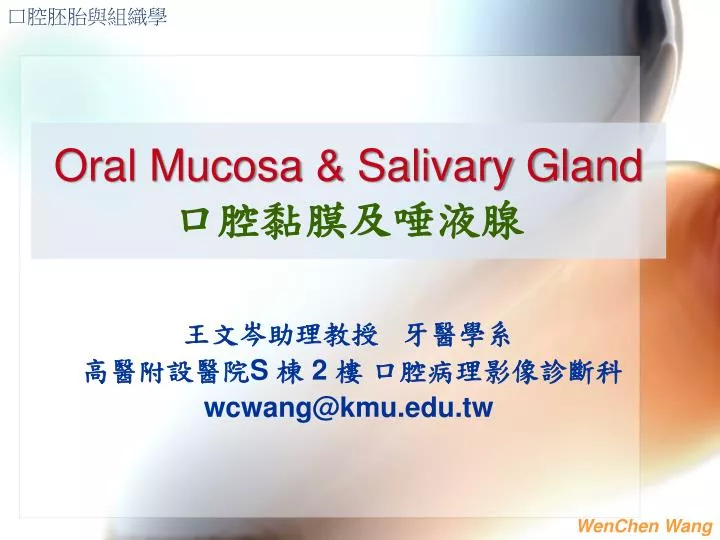
口腔胚胎與組織學. Oral Mucosa & Salivary Gland 口腔黏膜及唾液腺. 王文岑助理教授 牙醫學系 高醫附設醫院 S 棟 2 樓 口腔病理影像診斷科 wcwang@kmu.edu.tw . 學習重點 : 認識口腔各部位口腔黏膜、唾液腺之 : (1) 構造分類 (2) 組成 (3) 功能 學習資源及主要圖片引用 : 1.Nanci A: Ten Cate’s Oral Histology, Development, structure, and function 7th ed.

E N D
口腔胚胎與組織學 Oral Mucosa & Salivary Gland口腔黏膜及唾液腺 王文岑助理教授 牙醫學系 高醫附設醫院S 棟 2 樓 口腔病理影像診斷科 wcwang@kmu.edu.tw
學習重點: 認識口腔各部位口腔黏膜、唾液腺之: (1) 構造分類 (2) 組成 (3) 功能 學習資源及主要圖片引用: 1.NanciA: Ten Cate’s Oral Histology, Development, structure, and function 7th ed. 2.Orban BJ :Orban’s oral histology and embryology,9th ed. p.261-334 3.Avery JK: Essentials of Oral Histology and Embryology: A clinical approach. 2nd ed. p.164-83 4.高醫大 口腔病理診斷科
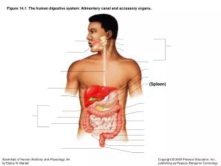
3 黏膜 MUCOUS MEMBRANE Definition: -Moist lining of the intestinal tract, nasal passages and other body cavities that communicate with the exterior Oral mucosa: Oral mucous membrane Ref. 1
各部位的口腔黏膜 正常口腔黏膜除舌背及硬顎外, 均為平滑外觀, 粉紅到桃紅色表面。 上下唇- • 唇紅緣(vermillion border) • 唇黏膜(labial mucosa) • 唇繫帶(labial frenum) • 前庭區(vestibule) Ref. 4
頰- • 頰黏膜 (buccal mucosa) • 咬合線(occlusal line, or linear alba) • 腮腺開口(orifice of Stenson’s duct) Ref. 4
Fordyce’s granule: 異位的皮脂腺 Ref. 4
嚼食檳榔者的黏膜 Ref. 4
舌背 舌- 口底- 舌背 唾液腺開口 (dorsal surface) 舌腹 (ventral surface) 舌繫帶 (lingual frenum) 舌側緣 (tongue border) 舌腹 口底 舌繫帶 Ref. 4
舌繫帶過短 (Tongue Tie) Ref. 4
牙齦 游離牙齦 Free ginigva 齒間乳突 (interdental papilla) 固著牙齦 (Attached gingiva) 齒槽黏膜 (alveolar mucosa ) 前庭區 (Vestibulae) Ref. 4
病態牙齦 Ref. 4
硬顎 顎部- 硬顎(hard palate) 軟顎(soft palate) 懸雍垂(uvula) 軟顎 Ref. 4
口腔黏膜的功能FUNCTIONS OF ORAL MUCOSA • 保護 Protection • 感覺 Sensation • 分泌 Secretion • 溫度調節 Thermal regulation
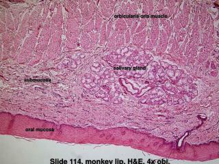
黏膜的基本構造 1.表皮(epithelium).....表皮 表皮細胞、神經末稍 角化上皮與非角化上皮 2.固有層(lamina propria)..真皮 膠原纖維、微血管及神經等結 締組織 3.黏膜下組織(submucous layer).. ...........皮下組織 小血管、神經、腺體(唾液腺和皮 脂腺) Ref. 1
Oral mucoperiosteum Ref. 1
EPITHELIUM表皮或上皮 ..複層鱗狀細胞 stratified squamous cell 角化 Keratinized 未角化 Nonkeratinized基底層 basal basal棘狀層prickle prickle顆粒層granular 中間層intermediate角質層keratinized 表層 superficial Ref. 1
上皮的角化過程Keratinization Basal cell prickle granular keratinized layer D.角質細胞 C.顆粒細胞 B.棘狀細胞 Skin… 52-75 days Gut… 4-14 days Gingiva… 41-57days Cheek… 25days A.基底細胞 Ref. 3
Cuboid or columnar shape Containing bundles of tonofibrils and cell organs Synthesize DNA, protein Most cell divisions Basal cell layer Ref. 3
Irregularly polyhedral shape Conspicuous tonofibril bundles Membrane-coating granules in upper part Intercellular bridge- Desmosome Most active in protein synthesis Prickle cell layer (spinous cell layer) Ref. 3
Flatter & wider Keratohyaline granules Tonofibrils Membrane-coating granules Synthesis protein Granular cell layer Ref. 3
Keratin layer • Flattened & dehydrated • Lost all organelles • Cell filled with packed fibrillar material orthokeratin parakeratin Ref. 1
Intermediate layer of nonkeratinizedepi. • Flattened cells • Tonofilaments & glycogen Superficial layer of nonkeratinizedepi. • Flattened cells • Tonofilaments & glycogen • Fewer organells • Nuclei persist Ref. 1
Melanocyte melanin, premelanosome, melanosome • Langerhan’s cell regulatory, antigen trap • Merkel’s cell tactile sensory • Lymphocyte inflammatory response **Clear cells: Melanocyte, Langerhan’s cell, Merkel’s cell Nonkeratinocytes in the oral epi. Ref. 1
Desmosome (Hemidesmosome) Tight junction Gap junction Intercellular Junctions Ref. 1 Intercelluar bridge
FUNCTIONAL CLASSIFICATION OF ORAL MUCOSA口腔黏膜之功能性分類 • 角化性上皮 • 咀嚼性黏膜-硬顎及牙齦 • 唇紅緣 • 非角化性上皮(襯裡性上皮) • 唇黏膜、齒槽黏膜、前庭區 • 特化上皮 • 舌背
1.different zones of palatine mucosa • gingival region, palatine raphae, fatty zone, glandular zone(mucous gland) Hard Palate Ref. 3
2. tight fixed to the underlying periosteum * No submucous layer: gingiva region and palatin raphe (mucoperiosteum) 3. epithelium uinform in form, well-keratinized Ref. 1
1. Parakeratinized 75%, orthokeratinized 15%, nonkeratinized 10% 2. often showing stippled surface Gingiva Ref. 3
3. Free gingiva, free gingiva groove, attached gingiva, interdentalgingiva, mucogingival junction Ref. 1,3
4. col depressed part of interdental papilla, thin nonkeratinizedepi. Ref. 1
Transitional zone between skin of lip and lip mucosa No gland Vermilion border of lip Ref. 1,4
… Lining or reflecting mucosa • lip,cheek, alveolar mucosa, vestibular fornix, mouth floor, soft palate, ventrum of tongue • Thick nonkeratinized epi. and varied amount of loose textured submucosa (containing fat & gland) movably attached to the deep structure Nonkeratinized Areas Ref. 2
特化上皮- 舌背 …Dorsal lingual mucosa 舌乳頭 (Lingual Papillae) 1.絲狀乳頭 Filiform papilla: 舌前方全部, 無味蕾 2.蕈狀乳頭 Fungiform papilla: 舌尖 3.葉狀乳頭 Foliate papilla: 舌側緣 4.輪廓狀乳頭 Circumvallate papilla: 舌根處, 8-10個 Ref. 1
Oral mucosa Circumvallate papilla Ref.1 Ref.4
Oral mucosa Ref.1
Taste Buds 味蕾 1. Ovoid or barrel-shaped intraepi. organ 2. Taste pore 3. Supporting cells 4. 10-12 neuroepithelial cells 神經上皮細胞 (receptors of taste味覺受器) Ref. 1 Ref. 3
味覺 • Sweet-fungiform papilla • Salty-fungiform • Bitter -circumvallate • Sour- foliate 舌的味覺控制 舌前三分之二:顏面神經 舌後三分之一:舌咽神經 Ref. 3
ORAL MUCOSA • Structure of oral mucosa Keratinized vs. Nonkeratinized Keratinocytes in the oral epi Nonkeratinocytesin the oral epi • Functional classification Keratinized areas Nonkeratinized areas Specialized mucosa Summary
SALIVARY GLANDS 唾液腺
唾液腺的分類CLASSIFICATION OF SALIVARY GLANDS 1.以部位 2.以大小 主唾液腺Major Salivary Gland • 耳下(腮)腺 Parotid gland • 頷下腺 Submandibular gl. • 舌下腺 Sublingual gland 小唾液腺Minor Salivary Gland • 唇、頰、舌、顎…Labial and buccal, palatine, lingual
唾液腺的分類CLASSIFICATION OF SALIVARY GLANDS 3. 以構造 • 黏液腺 Mucous gland-潤滑作用為主 • 漿液腺 Serous gland-含較多消化酶 • 混合腺 Mixed gland Ref. 1
唾液腺的構造STRUCTURE OF SALIVARY GLANDS • 腺泡細胞 Acinic cells • 管道系統 Duct system • 結締組織 Connective tissue: • 纖維間隔及被膜 fibrous septa and capsule • 血管 blood vessels • 神經 nerves Ref. 1
acinic cells mucous cell , serous cell or mixed acini Terminal tubule: acinic cell + myoepithelial cell Terminal secretory units: Terminal tubule+ intercalated duct cell Ref. 1
Mucous cell • pale • Low-protein, high carbohydrate • Mucin: glycoprotein, sialic acid • Viscous • Lubrication Ref. 3
Serous cell • Dark stain • High -protein, low carbohydrate • rER, lysosome, mitochondria, secretory granule, zymogen granules (amylase) • Watery consistency • Digestion Ref. 1, 3
Myoepithelial cell • Surrounding the acinic cell and intercalated duct • Long process • like smooth m. in ultrastructure (含肌動蛋白actin) • Contractile function, helping to extraction Ref. 1
Demilunes: mixed acini mucous cells (inner) + serous cells • Cell Junctions: • Tight junction • Intermediate junction • Desmosome characteristics of ectodemal origin Ref. 1