Download
1 / 49
610 likes | 1.25k Views
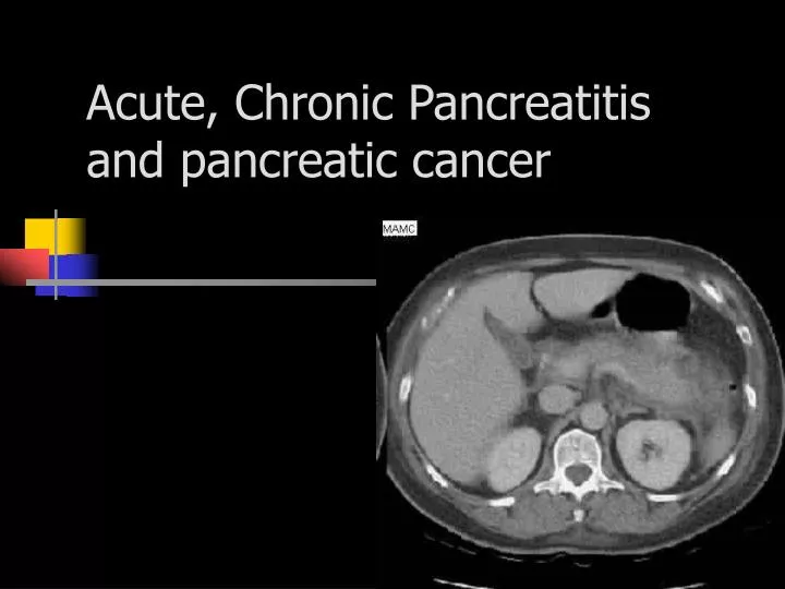
Acute, Chronic Pancreatitis and pancreatic cancer. Pathophysiology of acute pancreatitis. Triggering mechanism not exactly understood

E N D
Pathophysiology of acute pancreatitis • Triggering mechanism not exactly understood • When pancreas becomes damaged or ducts become blocked, trypsin inhibitor accumulates and activates pancreatic secretions that escape into the surrounding tissue, resulting in inflammation, thereby causing acute pancreatitis
Pathophysiology • Release of kallikrein and chymotrypsin results in increased capillary membrane permeability, leading to leakage of fluid into the interstitium and development of edema and relative hypovolemia • Elastase is the most harmful in terms of direct cell damage, it causes dissolution of the elastic fibers of blood vessels leading to hemorrhage • Phospholipase A in the presence of bile, destroys phospholipids of cell membranes causing severe pancreatic and adipose tissue necrosis • Lipase flows into damaged tissue and is absorbed into systemic circulation, resulting in fat necrosis of the pancreas and surrounding tissues
Edematous (Interstitial) Pancreatitis • Usually mild • Resolves in about 7 days • Results in fluid accumulation and swelling
Severe or Necrotizing Pancreatitis • Associated with a high degree of complications and mortality • Caused by the release of cytokines and other proinflammatory mediators that produce a hyperinflammatory reaction, resulting in cell death and tissue damage
Acute Pancreatitis: Damage and Destruction • If pancreatitis damages the islets of Langerhans, diabetes mellitus may result • Severe sudden pancreatitis may cause diabetic acidosis, shock and coma
Alcohol abuse} Gallstones } both 80% Mechanical/structural injury Sphincter of Oddi dysfunction Blunt trauma Post-ERCP 5% Pancreatic malignancy PUD ( penetrating) Medications Azathiprine Sulfonamides Furosemide Metabolic Hypertriglyceridemia Hypercalcemia Infectious Viral (mumps, rubella, CMV) Idiopathic10% Causes of Acute Pancreatitis
Clinical Presentation • Epigastric pain rapidly increasing in severity • Diffuse abdominal pain with radiation to back • Pain rarely only in left upper quadrant • Restlessness • Prefer to sit and lean • Nausea/Vomiting • Fever • Tachycardia
Abdominal Examination • Decreased or absent bowel sounds • Abdominal tenderness • Guarding • Palpable mass in epigastric area • Jaundice if there’s obstruction of the bile duct • Cullen’s sign • Grey Turner’s sign
Clinical Manifestations • Abdominal distention • Abdominal guarding • Abdominal tympany • Hypoactive bowel sounds • Severe disease: peritoneal signs, ascites, jaundice, palpable abdominal mass, Grey Turner’s sign, Cullen’s sign, and signs of hypovolemic shock
Investigation • Serum amylase raises within hours and remains for 4-5 d ( 3-5 x normal) • Urine amylase increased for 1-2 weeks • Elevated WBC • Decreased serum calcium • Elevated serum bilirubin, AST, ALT, LDH, and alkaline phosphatase • Abdominal XRAYS and CT’s showing pleural effusions and bowel dilation and ileus • Serum triglycerides >150mg/dl
Amylase • levels greater than three times the upper limit is needed for dx ( isoamylase is more specific) • Serum amylase levels may be normal if due to alcohol abuse or hypertriglyceridemia
Abdominal Source Biliary obstruction Bowel obstruction Perforated ulcer Appendicitis Mesenteric ischemia Peritonitis Salivary Parotitis Unknown Source Renal failure Head trauma Burns Postoperative DKA Other causes of Amylase Elevation
Diagnostic Evaluation • Serum lipase is more sensitive and specific • An elevation of greater than three times the normal value usually confirms acute pancreatitis • A lipase-to-amylase ratio of greater than 2 is usually evident with pancreatitis related to alcohol abuse
Imaging • Plain abdominal x-rays for visualizing gallstones or a gas-filled transverse colon ending at the area of pancreatic inflammation • colon cut-off sign • Abdominal ultrasound • Cholelithiasis, biliary sludge, bile duct dilation, and pseudocysts • CT of abdomen • MRCP • EUS
Ranson’s criteria of severity At admission: • Age > 55 • WBC > 16,000 • Glucose > 200 • LDH > 2x normal • ALT > 6x normal During initial 48 hr: • PaO2 < 60 mmHg • B.Urea ↑ >5 mg/dl • S. Ca < 8 mg/dl • ↓ in PCV > 10% • Base deficit > 4mEq/l • Estimated fluid sequestration > 600 ml Mortality: 1% if <3 scores, 10-20% if 3-5, > 50% if ≥ 6
Complications – Local • Necrosis • Sterile • Infected - abscess • Pseudocyst • Ascites • Intraperitoneal hemorrhage • Thrombosis • Bowel infarction • Obstructive jaundice
Pulmonary Pleural effusions Atelectasis Mediastinal abscess ARDS Cardiovascular Hypotension Sudden death Pericardial effusion Hematologic DIC Gastrointestinal PUD Erosive gastritis Blood vessel erosion Portal vein thrombosis Renal ARF Renal artery/vein thrombosis Complications – Systemic
Management • Fluid Management • Nutritional support • Rest gut • TPN • Pain management • Supporting other organ systems
Treatment • IV replacement of fluids, proteins, electrolytes and blood • Withholding food and fluids to rest the pancreas • NG tube suctioning • Drugs • Peritoneal lavage • Surgical drainage • Laparotomy to remove obstruction
Fluid Resuscitation • Patients with acute pancreatitis may have fluid shifts of 4 to 12 L into retroperitoneal space and peritoneal cavity due to inflammation • In severe acute pancreatitis, blood vessels in and around the pancreas may also become disrupted, resulting in hemorrhage. • Replace fluids with colloids, crystalloids, or blood products • Monitor for S&S of hemorrhage ( chart)
Rest the Pancreas • NPO status • Resting the gut is recommended until the patient no longer reports abdominal pain and serum amylase has returned to normal • Provide nutrition enterally using a jejunal tube to prevent pancreatic enzyme secretion. • If parenteral therapy is used, the solution is usually a mixture of hypertonic glucose and amino acids. • The use of lipid emulsion is contraindicated during acute phase because it increases pancreatic exocrine secretion.
Pharmaceutical Treatment • Pethidine for pain • Morphine causes spasm of Sphincter of Oddi • Pneumatic compressions
Management - Antibiotics • Infected necrosis has significant mortality (40%) • prophylactic antibiotics in severe pancreatitis with necrosis • Imipenem, cefuroxime, ceftazidime + amikacin + metro, Ofloxacin + metro, cipro + metro your choice • If used, it should be given no longer than 7 to 14 days • Gut decontamination is not recommended
Goals for Treatment • Supportive care • Pain control • Antiemetics • IVF • NPO • Lessening inflammation and necrosis • ERCP with stone extraction or stent placement • Treating complications • Preventing recurrence
ERCP IN ACUTE PANCREATITIS Emergency ERCP with biliary sphincterotomy and stone extraction when stones are identified in the common bile duct improves outcome in severe acute pancreatitis. Greatest benefit occurs in those patients who have ascending cholangitis
Prognosis • ~90% mild, self-limited • Usually resolves in 3-7 days • ~10% severe requiring ICU admission • Mortality may approach 50% in severe cases
Chronic pancreatitis • Pathophys - irreversible parenchymal destruction leading to pancreatic dysfunction • most frequent in men with peak incidence between the age 35 and 45 years. • Weight loss is multifactorial: maldigestion, fear of eating, anorexia, nausea, and vomiting • Constipation, flatulence • Steatorrhea- does not occur until 90% of pancreatic function is lost. • Diabetes- when more than 80% of the gland is destroyed.
Clinical picture • Abdominal pain occurs in 50% to 80% , pain is dull or boring in quality and worsens after eating. The pain is located in the epigastric area and often radiates to the back. Two patterns of pain • Short, relapsing episodes lasting days to weeks, separated by pain-free intervals. • Prolonged, severe, unrelenting pain.
Chronic pancreatitis • #1- etiology is chronic alcohol abuse (90%) • Gallstones • Hyperparathyroidism • Congenital malformation (pancreas divisum) • Idiopathic • MRCP of pancreas divisum
Evaluation • or normal amylase and lipase • Plain AXR / CT may show calcified pancreas • Pain management critical • Alcohol cessation may improve pain • Narcotic dependency is common
ERCP • ERCP is a highly sensitive radiographic test for CP.
CT features • The cardinal CT features of CP are pancreatic atrophy, calcifications, and main pancreatic duct dilation .
Direct PFTs require placement of double-lumen, gastroduodenal tubes for pancreatic fluid collection after intravenous CCK or secretin stimulation. Direct tests are sensitive for early CP.
TREATMENT(1) Pain relief — • Avoiding alcohol • Modifying meals • Non-narcotic analgesics : NSAIDs such as ibuprofen • Pancreatic enzyme supplements • Narcotic analgesics
TREATMENT(2) Treatment of fat malabsorption and steatorrhea — • Reducing fat intake • Lipase supplements • Medium chain triglycerides
INTERVENTION IN CHRONIC PANCREATITIS • Endoscopic therapy • Dilatation or stenting of pancreatic duct strictures • Removal of calculi (mechanical or shock-wave lithotripsy)
Intervention… • Surgical methods • Partial pancreatic resection, preserving the duodenum • Pancreatico-jejunostomy
Complications • Exocrine insufficiency typically manifests as wt loss and steatorrhea • If steatorrhea present, a trypsinogen level < 10 is diagnostic for chronic pancreatitis • Manage with low-fat diet and pancreatic enzyme supplements (Pancrease, Creon) • Endocrine insufficiency - from islet cell destruction which leads to diabetes
Incidence • One of the leading causes of cancer mortality • Incidence increases with age • Rarely before 50 y/o – usually 60 to 70 y/o • Slightly more in men • Less than 20% live longer than one year
Etiology of Pancreatic Cancer • Exact cause unknown • Smokers at high risk • High fat, high protein, high caffeine, high alcohol diets • May be genetic
Pathophysiology • Usually arises from epithelial cells of the pancreatic ducts • Tumor discovered in late stages so has spread throughout pancreas • Can be a result of metastatic disease from lung, breast, thyroid, kidney, or skin • Rapid growing with spread to surrounding tissue
More Patho • Most common site is the head of the pancreas • Necrotic pancreatic tumors increase thromboplastic factors • Thrombophlebitis seen as a result
Diagnostics • No specific blood tests to diagnose • Elevated amylase, lipase, alkaline phosphatase, bilirubin, CEA, C19-9 • CT, Ultrasonography • Needle biopsy • Paracentesis • ERCP – most definitive diagnostic test
Signs and Symptoms • Vague, dull, abdominal pain • Weight loss, weakness • Anorexia, nausea, vomiting • Glucose intolerance • Flatulence • GI bleeding • Ascites • Leg DVT • Jaundice (if head of pancreas involved) • Clay colored stools • Dark urine
Clinical Management • Goal is to prevent spread of tumor • Chemotherapy or radiation • Pain control • Total resection • Head of pancreas removal • Whipple procedure
Whipple Procedure • Radical pancreaticoduodenectomy • Used for cancer of the pancreas head only • Removal of: • Pancreas head • Duodenum • Stomach • Portion of jejunum • Gallbladder • Spleen • Duct anastomoses: pancreatic& common bile to jejunum
Clinical Management Continued • Nutritional support: TPN, tube feeding • Needs critical care nursing postoperatively • Fluid and electrolyte replacement • Glucose monitoring