Download
1 / 38
420 likes | 1.91k Views
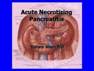
Acute & Chronic Pancreatitis. 11/01/2005 Chp. 87 Tintinalli Bogdan Irimies D.O. Acute Pancreatitis: Epidemiology. Clinical presentation can vary from mild abdominal pain to refractory shock 90% of acute pancreatitis is secondary to acute cholelithiasis or ETOH abuse

E N D
Acute & Chronic Pancreatitis • 11/01/2005 • Chp. 87 Tintinalli • Bogdan Irimies D.O.
Acute Pancreatitis: Epidemiology • Clinical presentation can vary from mild abdominal pain to refractory shock • 90% of acute pancreatitis is secondary to acute cholelithiasis or ETOH abuse • List if causes is extensive: Cholelithiasis, ETOH, drugs, infection, inflammation, trauma, metabolic disturbances
Drug Induced Pancreatitis • Drugs assoc. w/pancreatitis: • Amiodarone, amlodipine • Antibiotics(macrolides,sulfa, FQ’s, rifampin) • Antiepileptics (carbamazepine, valproic acid, topiramate) • Hyperlipidemic drugs • Antineoplastic agents • Antipsychotics (risperdal)
Drug Induced Pancreatitis • Drugs cont’d: • Antiretrovirals: all types • Diuretics • GI agents: H2 blockers, PPI’s • Glucocorticoids • NSAIDS • ASA
Pathophysiology • Central cause appears to be activation of the digestive zymogens in the pancreatic acinar cells and subsequent autodigestion of the pancreas. • Number of factors(endotoxins, toxins, ischemia, infections, anoxia) trigger activation of proenzymes
Pathophysiology • Activated proteolytic enzymes such as trypsin digest cellular membranes within pancreas and cause edema, interstitial hemorrhage, vascular damage, coagulation and cellular necrosis. • This can lead to extension of localized process into generalized systemic inflammatory response • Can lead to shock, ARDS, Multi-organ system failure
Clinical Features • Major symptom is midepigastric or left upper quadrant pain: described as constant, boring pain that radiates to back, flanks, chest or lower abdomen. • Nausea/vomiting or abdominal bloating • PE: low grade fevers, tachycardia, +/- hypotension
Clinical Features • Respiratory symptoms: atelectasis, pleural effusion, ARDS • Abdominal exam: epigastric tenderness, peritonitis, Cullen sign(bluish discoloration around umbilicus), Grey Turner sign (bluish discoloration of flanks) • Pts. May present in hypovolemic shock and MOSF • Hypotension secondary to 3rd spacing, hemorrhage, increased vascular permeability, vasodilation, cardiac depression, vomiting
Diagnosis • Amylase: found in pancreas & salivary glands • Low levels found in many tissues so this test is nonspecific • Amylase may be even normal in acute pancreatitis • Poor specificity
Diagnosis • Lipase: found predominantly in pancreas but also in gastric, intestinal mucosa and liver • Cleared by the kidney so renal failure will elevate levels • Most appropriate cut-off is 2-3 x normal level • More accurate test than amylase, better specificity (90% vs. 75%)
Diagnosis • Xrays of chest/abdomen: useful for r/o other diagnosis. • Calcification of pancreas seen in chronic pancreatitis • May see sentinel loop, elevated hemi-diaphragm, pleural effusion • U/S may detect gallstones • CT best study for grading severity if disease, prognosis.
Diagnosis • Prognostic markers: Ranson criteria predicts pt. outcome • Age >55 • BS >200 • WBC >16,000 • AST >250 • LDH >700 • Features portend a worse prognosis, but they have poor predictive value in acute setting and does not improve clinical judgment
Diagnosis • CT of abdomen: • Estimates severity and prognosis • Complications include phlegmons, abscesses or pseudocysts. • Usually seen 2-3 weeks after acute pancreatitis
Complications of Acute Pancreatitis • Pulmonary: pleural effusions, atelectasis, hypoxemia, ARDS • CV: myocardial depression, hemorrhage, hypovolemia • Metabolic: Hypocalcemia, hyperglycemia, Hyperlipidemia, coagulopathy/DIC • Others: Colonic perforation, ARF. Arthritis, pseudocyst, abscess
Treatment: • General principle: rest the pancreas • Fluid resuscitation • NG tube only if needed • Pain control, anti-emetics • ATBX only in severe disease • Cover polymicrobial, GNB • IV imipenem or quinolone in combination w/Flagyl
Disposition: • Pts. w/mild pancreatitis w/no evidence of systemic disease and low likelihood of biliary disease may be managed as outpts. if tolerating oral fluids and pain control is adequate • All others need to be admitted
Chronic Pancreatitis • Defined as chronic inflammatory condition that causes irreversible damage to pancreatic structure and function • Causes: ETOH abuse, malnutrition, hyperPTH, pancreas divisum, ampullary stenosis, cystic fibrosis, hereditary, trauma, idiopathic
Chronic Pancreatitis • Chronic pancreatitis results in interstitial inflammation w/duct obstruction and dilation leading to parenchymal loss and fibrosis. • Loss of both exocrine and endocrine • Clinicically significant malabsorption occurs when 90% of pancreas is lost.
Chronic Pancreatitis • Presents as midepigastric abdominal pain, nausea, vomiting • Pts. May appear chronically ill, w/sign of pancreatic insufficiency such as weight loss, steatorrhea, clubbing, polyuria • Differentiating acute vs chronic pancreatitis is difficult b/c primary distinction is based on disease reversibility
Chronic Pancreatitis • Amylase and lipase may be normal if pancreas is fibrotic • CT scan may help ID pseudocyst or abscess • Tx: IVF’s anti-emetics, narcotics • Pancreatic extracts to improve absorption and pain • If pain is increasing or intractable, image pancreas to look for complications
Disposition • Pts. Maybe discharged home if all the complications have been ruled out • Hospitalize if intractable pain.
Questions • 1. Which of the following are common causes of pancreatitis • A. infection • B. Gallstones • C. ETOH • D. Drugs • E. all of above
Questions • 2. Which of the following are complications of pancreatitis: • A. ARDS • B. Shock • C. pancreatic insufficiency • D. pleural effusions • E. all of above
Questions • 3. True or false: many meds can cause pancreatitis? • 4. True or false: Grey Turner and Cullens sign are signs of hemorrhagic pancreatitis? • 5. True or false: There is no single lab test that can reliably diagnose pancreatitis?
Answers • 1. E • 2. E • 3. T • 4.T • 5. T
Case of the Day: • HPI: 54 y/o WF presented to ER after being found on the ground s/p fall by her son. Pt. was found to be lethargic, weak, dizzy. Pt. had been vomiting the preceding 2-3 days. C/O diffuse abdominal pain. • ROS: + weight loss 50 lbs. over past year, rest of ROS neg.
Case of the Day • PMHx: 1. Anemia 2. GERD 3. HLD 4. Hypokalemia 5. Herniated disc • PSHX: 1. TAH 2. Chole • NKDA • Meds: Urocrit, Zyprexa, Prevacid, Vicodin • Soc Hx: Denies ETOH, + 1pk. Day smoker, no drugs • Fam Hx: N/C
Physical Exam • VS: 36.3, 96/60, 109, 14, 97% RA • Gen: A&o x1 , cachetic, difficult to arouse • ENT: mm dry, otherwise normal • CV: Tachy, S1,S2 no m/c/r • Pulm: LCTAx2 • GI: + BS, soft, diffuse TTP, No R/R/G • Rectal: heme + stools (done by Dr. Holencik) • Neuro: intact, no focal deficits • Ext: good pulses, no edema
Labs • EKG: ST 109 bpm • CBC: WBC 8.5 10.7/32.4 Plt 440 MCV 102.2, Fe+ def. anemia • CMP: Na 143, K 3.6, Cl 109 CO2 12, GLU 63 BUN 17 Cr. 1.0 Alb. 3.3 AST/ALT nml, Amylase,lipase normal Mg 1.8 • CPP neg. x 1, CXR: NAD CT head: neg.
Labs • UA: 1+ protein, 2+ blood • ABG: 7.24/26/95/10/96% RA • APAP/ASA neg. • UDS: + BZD TSH 0.23 (L) L.A. 1.6 • ETOH 0.002 • Serum acetones: large amount • Serum Osm: 294
Methanol Uremia Dka Inh/iron Lactic acidosis Ethylene glycol ASA CO Cyanide AKA/starvation Tolulene D/Dx: mental status change/metabolic acidosis
Alcoholic ketoacidosis(AKA): • AKA is a wide anion gap metabolic acidosis often assoc. w/acute cessation of ETOH consumption after chronic ETOH abuse. • Key features are ingestion of large amounts of ETOH, relative starvation, volume depletion
AKA • Relative starvation, lack of glucose/glycogen stores, insulin deficiency, production of counter-regulatory hormones • Lipolysis promoted w/conversion of acetyl Co A to ketones
N/V abd. Pain Tachycardia & Tachypnea SOB Tremulousness Dizziness Hematemesis, melena Hepatomegaly Mental status change Seizure/syncope Muscle pain Fever Clinical Features:
Lab: • ETOH: low or none • Elevated anion gap caused by ketones • Serum ketones: maybe neg. or high (assay detects AcAc/acetone, BHB predominant ketone in AKA) • Electrolytes: hypophosphatemia, hypokalemia, hyponatremia, hypoglycemia • Acid Base: maybe mixed met. Acidosis & met. Alkalosis(vomiting, volume depletion)
Treatment: • Glucose administration to promote insulin secretion • IVF: D5NS , HCO3 if pH<7.1 • Thiamine • Admit for acidosis
Hyperkalemia: EKG see III-19 • Tall, tenting of T-waves • Prolongation of QRS & P-R interval • Low amplitude p-waves • AV blocks • Sine wave, V. Fib, asystole
Hypokalemia: see III-20 • Flattening of T-waves, U waves present • ST-depression • T-wave inversion • Advanced: PAT w/block