Download
1 / 27
500 likes | 2k Views
LECTURE 2:. Prokaryotic Cell Structure and Function. Microbiology and Virology; 3 Credit hours Atta- ur - Rahman School of Applied Biosciences (ASAB) National University of Sciences and Technology (NUST). Bacterial Cell Wall.

E N D
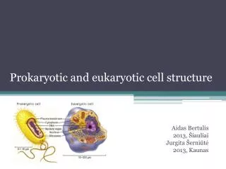
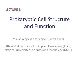
LECTURE 2: Prokaryotic Cell Structure and Function Microbiology and Virology; 3 Credit hours Atta-ur-Rahman School of Applied Biosciences (ASAB) National University of Sciences and Technology (NUST)
Bacterial Cell Wall • The cell wall is the layer, usually fairly rigid, that lies just outside the plasma membrane. • After Christian Gram developed the Gram stain in 1884, it soon became evident that most bacteria could be divided into two major groups based on their response to the Gram-stain procedure • Gram Positive Bacteria • Gram Negative Bacteria
Bacterial Cell Wall • All the structures from the plasma membrane outward, the Cell envelope • This includes the plasma membrane, cell wall and structures like capsules if present. • Space between the plasma membrane and the outer membrane; Peri-plasmic space.
Peptidoglycan Structure • Peptidoglycan (aka murein) is a mesh-like polymer composed of many identical subunits. • The polymer contains two sugar derivatives, • N-acetyl-glucosamine • N-acetyl-muramicacid • Several different amino acids
Peptidoglycan Structure In order to make a strong, mesh-like polymer, chains of linked peptidoglycan subunits must be joined by cross-links between the peptides. Often the carboxyl group of the terminal D-alanine is connected directly to the amino group of di-aminopimelicacid, but a peptide interbridge may be used instead. Most gram-negative cell wall peptidoglycan lacks the peptide interbridge. This cross-linking results in an enormous peptidogly- can sac that is actually one dense, interconnected network .
Peptidoglycan Structure, Teichoic Acids • Gram positive cell walls usually contain large amounts of teichoic acids, polymers of glycerol or ribitol joined by phosphate groups • The teichoic acids are covalently connected to either the peptidoglycan itself or to plasma membrane lipids; in the latter case they are called lipo-teichoicacids. • They are negatively charged, help give the gram-positive cell wall its negative charge. • They are important in maintaining the structure of the wall. • Teichoicacids are not present in gram negative bacteria.
Teichoic Acid Structure The segment of a teichoicacid made of phosphate, glycerol, and a side chain, R. R may represent D-alanine, glucose, or other molecules.
Peri-plasmicSpace of Gram-Positive Bacteria • The substance that occupies the periplasmic space is the periplasm. • The periplasm has relatively few proteins; this is probably because the peptidoglycan sac is porous and any proteins secreted by the cell usually pass through it. • Enzymes secreted by gram-positive bacteria are called exoenzymes. • Serve to degrade polymeric nutrients
Gram-Negative Cell Walls • Gram-negative cell walls are much more complex than gram-positive walls. • The periplasmic space of gram-negative bacteria is also strikingly different • It ranges in size from 1 nm to as great as 71 nm • It may constitute about 20 to 40% of the total cell volume • Nutrient acquisition—for example, hydrolytic enzymes
Gram-Negative Cell Walls • Some periplasmic proteins are involved in energy conservation • The denitrifying bacteria, which convert nitrate to nitrogen gas, and bacteria that use inorganic molecules as energy sources (chemolithotrophs) have electron transport proteins in their periplasm. • Other periplasmicproteins are involved in peptidoglycan synthesis and the modification of toxic compounds that could harm the cell.
Lipopolysaccharides (LPSs) • Outer to the peptidoglycan layer is lipopolysaccharides (LPSs) • LPS contain both lipid and carbohydrate, and consist of three parts: • lipid A • the core polysaccharide • the O side chain
The lipid A region contains two glucosamine sugar derivatives, each with three fatty acids and phosphate or pyrophosphate attached. • The fatty acids attach the lipid A to the outer membrane, while the remainder of the LPS molecule projects from the surface. • The core polysaccharide is joined to lipid A.
Lipopolysaccharides (LPSs) • Peptidoglycan layer and LPS are joined in two ways • The first is by Braun’s lipoprotein, the most abundant protein in the outer membrane. • This small lipoprotein is covalently joined to the underlying peptidoglycan, and is embedded in the outer membrane by its hydrophobic end. • The second linking mechanism involves the many adhesion sites joining the outer membrane and the plasma membrane.
Functions of Lipopolysaccharides (LPS) • The core polysaccharide usually contains charged sugars and phosphate, LPS contributes to the negative charge on the bacterial surface. • Bacterial attachment to surfaces and biofilm formation. • It aids in creating a permeability barrier to restrict the entry of bile salts, antibiotics, and other toxic substances that might kill or injure the bacterium. • The O side chain of LPS is also called the O antigen because it elicits an immune response. • G negative bacteria are able to rapidly change the antigenic nature of their O side chains, thus escaping host defenses.
GRAM POSITIVE Lipoteichoic acid Peptidoglycan-teichoic acid Cytoplasmic membrane Cytoplasm GRAM NEGATIVE Lipopolysaccharide Porin Outer Membrane Braun lipoprotein Periplasmic space Inner (cytoplasmic) membrane Cytoplasm
What happens in Gram staining? • Crystal Violet (purple) • Primary stain; positive stain. • Stains all cell walls purple. • Iodine • Mordant (Any substance used to facilitate the fixing of a dye to a fibre). • Combines with CV to form an insoluble complex that gets trapped in thicker peptidoglycan layers. • Ethanol • Decolorizer. • CV complex washed out of Gram negative organisms because it cannot be trapped by outer layer. • Safranin (pink) • Counterstain. • Stains all cells, but only the negative ones actually appear pink.
G -ve G +ve
ARCHAEAL CELL WALLS • Archaeal wall structure and chemistry differ from those of the Bacteria. • Archaeal cell walls lack peptidoglycan and also exhibit considerable variety in terms of their chemical make-up. Methanobacteriumformicicum, and (b)Thermoproteustenax. CW, cell wall; SL, surface layer; CM, cell membrane or plasma membrane; CPL, cytoplasm.
Gram Positive Type Archae • Their wall chemistry varies from species to species but usually consists of complex heteropolysaccharides. • Methanobacterium and some other methane-generating archaea (methanogens) have walls containing pseudomurein • A peptidoglycan like polymer that has L-amino acids instead of D-amino acids in its cross-links • N-acetyltal-osaminuronicacid instead of N-acetylmuramic acid, • β(1→3) glycosidic bonds instead of β(1→4) glycosidic bonds.
The Structure of Pseudomurein Archaeaβ(1→3) Glycosidic Bond Bacteria β(1→4) Glycosidic Bond
Gram Negative Type Archae • Many archaea that stain gram negative have a layer of glyco-protein or protein outside their plasma membrane • Some methanogens (Methanolobus), salt-loving archaea (Halobacterium), and extreme thermophiles (Sulfolobus, Thermoproteus, and Pyrodictium) have glycoproteins in their walls. • The layer may be as thick as 20 to 40 nm. • In contrast, other methanogens (Methanococcus, Methanomicrobium, and Methanogenium) and the extreme thermophile Desulfurococcus have protein walls.