Download
1 / 24
240 likes | 393 Views
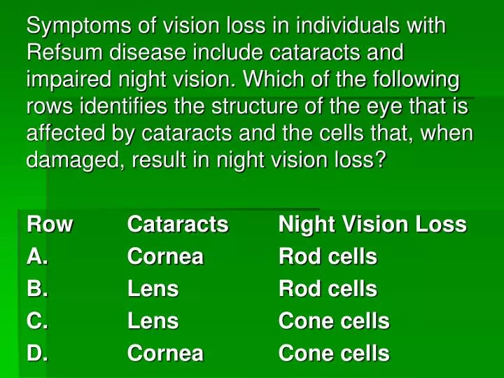
Symptoms of vision loss in individuals with Refsum disease include cataracts and impaired night vision. Which of the following rows identifies the structure of the eye that is affected by cataracts and the cells that, when damaged, result in night vision loss? Row Cataracts Night Vision Loss

E N D
Symptoms of vision loss in individuals with Refsum disease include cataracts and impaired night vision. Which of the following rows identifies the structure of the eye that is affected by cataracts and the cells that, when damaged, result in night vision loss? Row Cataracts Night Vision Loss A. Cornea Rod cells B. Lens Rod cells C. Lens Cone cells D. Cornea Cone cells
The Ear Chapter 14.3
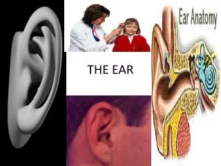
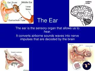
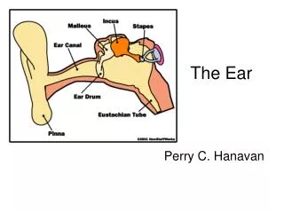
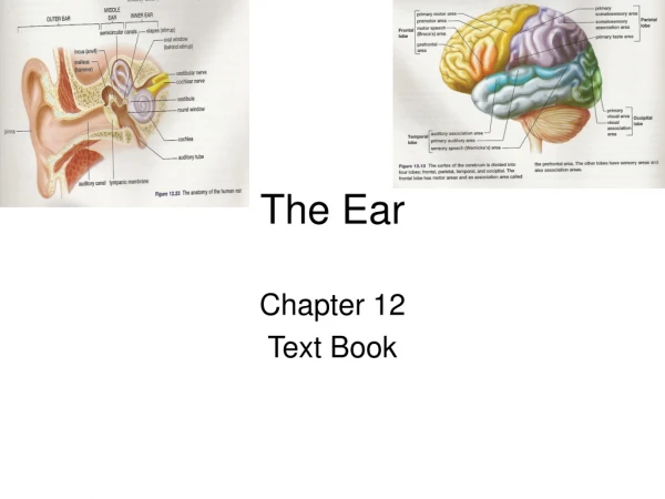
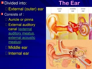
Structure of the Ear • When talking about the structure of the ear, we separate parts of the ear into three categories: • Outer Ear • Middle Ear • Inner Ear
Outer Ear • The outer ear is composed of two main parts: • The pinna which acts as a funnel to focus sound into: • The auditory canal which carries sound to the ear drum.
Middle Ear • The middle ear is composed of two parts: • The eardrum, which is known as the tympanic membrane. This takes the vibrations of sound waves and transfers it to: • The ossicles, which are small bones in the ear. These then continue the transfer of vibrations to the inner ear.
Ossicles • The ossicles are three bones: • The hammer (malleus). • The anvil (incus). • The stirrup (stapes)
Eustachian Tube • Sometime the pressure in the ear will be too high or too low for the inner ear. • To correct this, air will move through the eustachian tube to get to the right pressure. • The eustachian tube connects directly to your upper pharynx.
Inner Ear • The inner ear is made up of three structures: • The semicircular canals, which provide information about dynamic equilibrium. • The cochlea which contains audioreceptors. • The vestibule, which moves sound waves in and out of the cochlea as well as provide information about static equilibrium.
Hearing • Sound waves that reach the ear drum cause it to vibrate. • These vibrations are then passed onto the ossicles, which then magnify them. • The ossicles then pass this vibration onto the vestibule, through another membrane known as the oval window. • To prevent damage to the oval window, muscles in the ear will disconnect the stirrup from the oval window.
As the oval window is pushed in, another membrane called the round window gets pushed out. • This creates a wave of fluid through the cochlea, which it will interpret as sound.
Structure of the Cochlea • The cochlea is a coil of a single tube. • The tube is shaped as below with the listed structures.
Organ of Coti • The organ of Coti lies on top of the basilar membrane. • As the basilar membrane vibrates, hairs on the organ of Coti will make contact with a second membrane. • When this happens, it initiates an impulse in your audioreceptors.
Detecting Frequencies • The basilar membrane starts thicker at the beginning of the cochlea, and gets thinner as it goes. • Higher frequencies, which have higher energies, will cause the early membranes to vibrate. • Lower frequencies will cause the later membranes to vibrate. • Your brain will take where in the cochlea an impulse starts and translate that to a specific tone.
Equilibrium • Equilibrium is separated into two categories: • Static equilibrium, which relates to motion along one plane. • Dynamic equilibrium, which relates to 3 dimensional movement.
Static Equilibrium • As we learnt, the vestibule detects static equilibrium. • It contains two sacs: • The saccule which detects vertical motion (like the Spaceshot at Galaxyland). • The utricle, which detects head tilt.
These sacks are lined with cillia (little hairs) and filled with a gelatinous material containing tiny stones of calcium carbonate called otoliths. • When the sacks shift, the otoliths will move due to gravity. • When they do so, they brush the cillia, which triggers a nerve impulse. • This is then transmitted to the cerebellum for interpretation.
Dynamic Equilibrium • Dynamic equilibrium is detected by the semicircular canals. • They operate in much the same way as the vestibular sacs, but they respond to rotational movement.
Homework • Pg 458 1-2 • Pg 461 1-7, 10