Download
1 / 31
370 likes | 703 Views
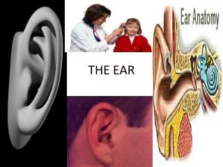
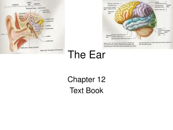
The Ear. Marlene Arceo Leonardo Cabrera Isaac Brabb Melanie Weyers. General functions of the ear. Two Main Functions : Assist with balance of individual Collect sounds and relay them to the brain. Outer ear. Structure : Made of tough cartilage covered by skin

E N D
The Ear Marlene Arceo Leonardo Cabrera Isaac Brabb Melanie Weyers
General functions of the ear • Two Main Functions: • Assist with balance of individual • Collect sounds and relay them to the brain
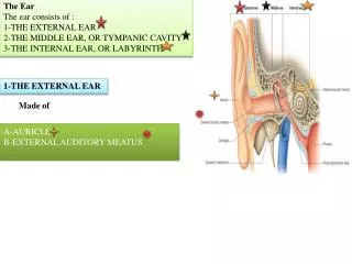
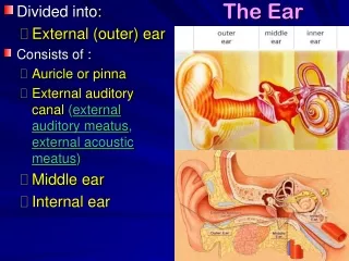
Outer ear • Structure: • Made of tough cartilage covered by skin • Contains external acoustic meatus • Secrets cerumen (protects ear canal) • Guarded by hair • Ends at the tympanic membrane (eardrum)
Outer ear (Cont.) • Function: • Auricle • Collects sound waves travelling through air • Directs sound waves into external acoustic meatus • Sound waves travel through tube all the way to the tympanic membrane • Tympanic Membrane • Semitransparent membrane covered by thin layer of skin on outside / mucous on inside • Oval margin / cone shaped • Moves back and forth in response to sound waves • Whatever sound wave enters the canal becomes processed and moves on to middle ear
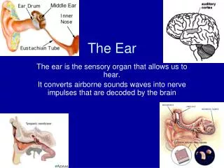
Middle ear -An air-filled, membrane lined space internal to the eardrum/tympanic membrane and external to the oval window of the cochlea -couples sound from air to the fluid via the oval window
Middle ear-Eustachian Tube • Links the nasopharynx to the middle ear • Remains closed under normal conditions 1. Pressure Equalization • When pressure inside middle ear is different form pressure of atmosphere, the tube can be opened from yawning, swallowing, or chewing because it lets small amounts of air and equalizes the pressures 2. Mucus Drainage • Eustachian tube allows fluid/mucus to drain out of the ear.
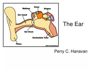
Middle Ear-OSSICLES • 3 small, movable bones • Vibrate when the eardrum/tympanic membrane vibrates from sound waves entering the ear canal • Hammer (malleus) • Bone connected to inside of the tympanic membrane and to the incus/anvil • Transmits sound vibrations to the incus • Anvil (incus) • Second bone connected to hammer to the stapes • Stirrup (stapes) • The third U-shaped bone connected to the stirrup and to cochlea • Smallest bone in the body
Muslces • Tympanic/Acoustic Reflex: muslces contract in response to loud sounds, thereby reducing the transmission of sound to the inner ear • Stapedius muscle-(smallest skeletal muscle in the body) connects to the stapes and is controlled by the facial nerve • Stapetensor tympanic muscle-connects to the base of the malleus and is under the control of the medial pterygoid nerve
Nerves • horizontal and chorda tympani branches of the facial nerve pass through the middle ear space • Damage to the horizontal branch during surgery can lead to partial mastoid process paralysis
Inner Ear • Location: • Within temporal bone • Functions: • Hearing and balance • Structures: • “Bony Labyrinth” • “Membranous Labyrinth” • Round Window • Eighth Cranial Nerve • Contains perilymphand Endolymph Fluid
Bony vs. Membranous Labyrinth • Membranous Labyrinth • Located INSIDE of the Bony Labyrinth • Smaller • Contains Endolymph • Ex: Inner tube of Bike Tire • Bony Labyrinth • Ducts created by the grooves and channels in the temporal bone • Larger • Contains Perilymph • Ex: Outer Tire of a Bike
Perilymph vs. Endolymph Fluid • Endolymph Fluid • Where actual perception of balance, equilibrium, and sound take place • Rich in Potassium • Located in Membranous Labyrinth • Perilymph Fluid • Similar composition to CSF • Rich in Sodium • Located in Bony Labyrinth • Shock buffer • Helps transmit waves to membranous labyrinth
Cochlea 1. Snail like structure for hearing 2. Converts sounds and vibrations from the external environment into electrical impulses 3. Transduction • When vibrations are made into impulses able to be interpreted by the brain • Vibrations stimulate oval window, causes the fluid to move through cochlea to the Organ of Corti • Sensory nerve “hair-like” receptors in the cochlea 4. Sensory cells in the cochlea perform transduction and the electrical impulse travels to the brain via Cochlear nerve
Semicircular Canals • Mainly for balance and steadiness • Contains Perilymph and Endolymph • When the body (especially the head) changes position, endolymph moves, hair cells in the canal sense the movement and relay the information to the brain. • Helps sense movement of the body and maintain balance
Vestibule • Regulates balance and equilibrium • Connects the semicircular valves and the cochlea • Also contains hair cells that sense changes in the waves and flow of Endolymph fluid to help maintain balance • Vestibular nerve relays information to regulate balance and equilibrium • Does not require conscience changes from the cerebrum • The body will automatically adjust itself • These help detect Lateral/Horizontal movement • Saccule: Inferior Portion of Vestibule • Utricle: Superior Portion of Vestibule
Round Window • Location: • Inner ear between the middle ear and the cochlea • Senses vibrations from middle ear, then indents into the inner ear to stimulate waves and movement of perilymph fluid. • Release to decrease pressure within the ear
Static equilibrium • Organs involved are located within utricle and saccule • Utricle and saccule contain hair cells that project vertically or horizontally depending between utricle and saccule • Hair contacts sheet of gelatinous material that ahs calcium carbonate crystals embedded • The hair cells have nerve fibers wrapped around their bases • Gravity stimulates hair cells to respond (i.e. bending head) • Gelatinous material sags in response to gravity • Stimulates hair cells, signal associated nerve fibers • Nerve impulses travel to the CNS • Informs brain of head’s position • Brain responds bye sending motor impulses to skeletal muscles which maintain balance
Dynamic equilibrium • 3 semicircular canals that are responsible for detecting changes in motion. • In each canal there is an ampulla • Communicates with utricle • Contains sensory organ crista ampullaris • Also contains sensory hair cells • When the head or torso moves, the semicircular canals move as well • This causes the hairs to bend, stimulating associated nerve fibers and as a result impulses travel to the brain.
Ménière’s Disease • Disease of the inner ear that causes vertigo (severe dizziness), ringing in the ears, hearing loss, and feeling of congestion in the ear. • Usually affects only one ear • Symptoms can be caused by: • The buildup of fluid in compartments of the ear (labyrinth) • It interferes with the normal balance and hearing signals between the inner ear and the brain • How is it diagnosed: • Medication to treat dizziness • Limit salt in diet / taking diuretics • Inject antibiotics (gentamicin) into the middle ear
Otitis • General term for infection or inflammation of the ear. • Classified according to whether it occurs suddenly for a short time or repeatedly over a long period of time • Otitis Externa • When it involved the outer ear and ear canal • Also called swimmers ear • Otitis Media • Involves the middle ear • Also called ear infection • Acute Ear Infection • Often painful • Starts suddenly / short period of time • Chronic Ear Infection • Ear infection does not go away / keeps reoccurring • Can cause long term ear damage
Works Cited Centers for Disease Control and Prevention. Centers for Disease Control and Prevention, 30 Sept. 2013. Web. 31 Mar. 2014. "Ears and Hearing." InnerBody. N.p., n.d. Web. 31 Mar. 2014. The Editors of Encyclopædia Britannica. "Vestibule (ear)." Encyclopedia Britannica Online. Encyclopedia Britannica, n.d. Web. 31 Mar. 2014. "Ménière's Disease." [NIDCD Health Information]. N.p., n.d. Web. 29 Mar. 2014. "Neuroscience for Kids - The Ear." Neuroscience for Kids - The Ear. N.p., n.d. Web. 31 Mar. 2014. "Special Senses II: The Ear." Special Senses II: The Ear. N.p., n.d. Web. 31 Mar. 2014. "X. The Organs of the Senses and the Common Integument. 1d. 4. The Internal Ear or Labyrinth. Gray, Henry. 1918. Anatomy of the Human Body." X. The Organs of the Senses and the Common Integument. 1d. 4. The Internal Ear or Labyrinth. Gray, Henry. 1918. Anatomy of the Human Body.N.p., n.d. Web. 31 Mar. 2014.