Download
1 / 29
290 likes | 525 Views
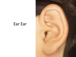
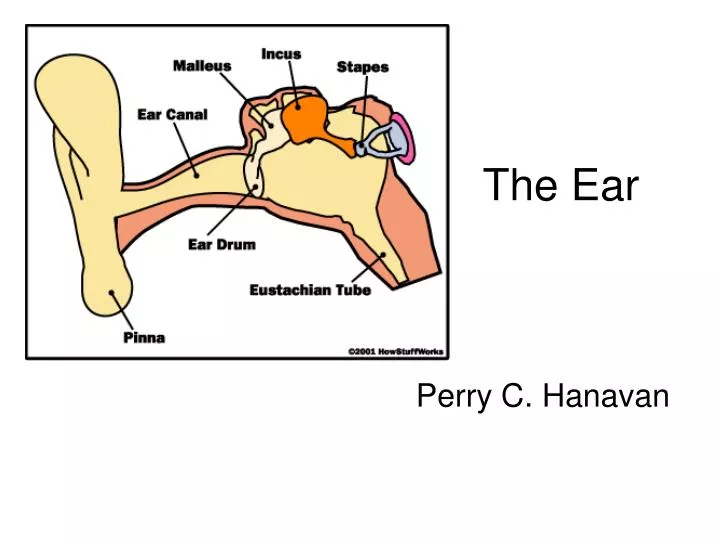
The Ear. Perry C. Hanavan. Outer Ear. Peripheral Outer ear Middle ear Inner ear Auditory nerve Central Brainstem Midbrain Cerebral. Peripheral. Outer Ear - Pinna. Middle Ear - Tympanum. Middle Ear – Tympanic Membrane. Middle Ear – Ossicles. Middle Ear - Ossicles.

E N D
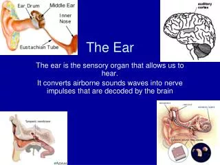
The Ear Perry C. Hanavan
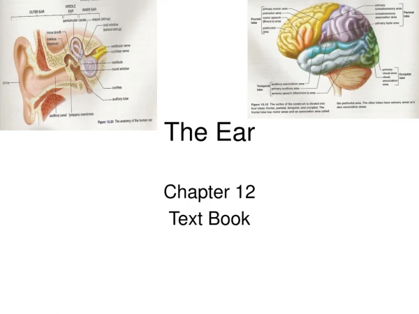
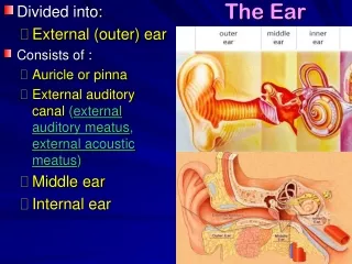
Outer Ear • Peripheral • Outer ear • Middle ear • Inner ear • Auditory nerve • Central • Brainstem • Midbrain • Cerebral
Basilar Membrane • Traveling wave
Inner Ear – OHC & IHC • IHC • Non-motile • Afferent • Responds to loud sounds • OHC • Motile • Efferent • Responds to soft sounds
Two Theories of Motility • Somatic prestin motor • Hair bundle motor
Tip-links –Hair Bundle Motor <<<IHC OHC >>>
Tip Link Structure • Tip Link Structure
2 Types of Auditory Neurons Paul Fuchs YouTube Paul Fuchs YouTube Type I Type II
Sup. Vest Nerve Standard ABR Facial Nerve Cross-section of Internal Auditory Canal HIGH- FREQUENCY FIBERS Acoustic Nerve Inf. Vest Nerve TUMOR The wave V latency used in the standard ABR IT5 and I-V delay measures is dominated by neural activity from the high-frequency regions of the cochlea. Thus, unless the tumor affects these high-frequency fibers sufficiently, standard ABR latencies will be normal. SmallTumor Abnormal Standard ABR Large Tumor Abnormal Standard ABR Small Tumor Normal Standard ABR