Download
1 / 1
10 likes | 124 Views
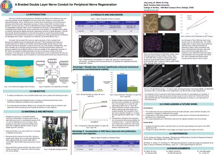
Jing Liang, Dr. Martin W. King, North Carolina State University College of Textiles, 1000 Main Campus Drive, Raleigh, 27606 jliang5@ncsu.edu. A Braided Double Layer Nerve Conduit for Peripheral Nerve Regeneration. 1.0 INTRODUCTION. 4.0 RESULTS AND DISCUSSION.

E N D
Jing Liang, Dr. Martin W. King, North Carolina State University College of Textiles, 1000 Main Campus Drive, Raleigh, 27606 jliang5@ncsu.edu A Braided Double Layer Nerve Conduit for Peripheral Nerve Regeneration 1.0 INTRODUCTION 4.0 RESULTS AND DISCUSSION Several hundred thousand people are affected by peripheral nerve defects every year, and many patients remain disabled for the rest of their lives. However, nerves have self-regenerating ability. Hence, artificial nerve conduits were introduced as a promising way to stimulate and enhance the regeneration process. A nerve conduit (NC) is a kind of nerve guidance channel (see Fig.1), which is fabricated into tubular shape by using degradable or non-degradable materials. When a NC is implanted into the body, it will be extended, compressed and rotated by the various movements of the patient. So, flexibility and an ability to maintain dimensional stability during the regeneration process is highly desirable. It should also provide directional guidance so the new nerve grows to reunite the severed stumps; meanwhile it should prevent the formation of scar tissue and retain growth factors secreted by the damaged nerve ends[1]. Properties of an ideal NC can be described in Fig. 2. Currently, Some known NCs have too rigid a structure or fail to maintain their dimensional stability during the regeneration process[2]. Micro-braiding technology is a versatile approach to obtain a soft, flexible and semipermeable tubular structure, with improved mechanical properties. However, there are only a few studies of braided NCs, and they invariably use a one layer structure with poor mechanical performance. Moreover, previous research has studied the effect of only one variable on the regeneration process. A NC which can integrate multiple parameters into the one structure is likely to be more successful. This study aims to design design and fabricate a multi-compartments NC that can combine multiple therapies, and to evaluate its mechanical performance and cell performance. Table 1. Basic Properties of Nerve Conduits a Fig 9. Schwann Cell Proliferation by MTT Assay a b b As it showed in Fig 8, 4DG has much more surface area for cell attachment. The LSCM images after 14 days culture also proved that 4DG had more and well stretched cell attaching to fibers, while round fibers had fewer cell attachment and some cells (red) were found to float outside fibers. As it showed in Fig 9, 4DG-2NC had much higher cell proliferation than round-2NC and hollow NC, which indicated that 4DG provided enough surface area for cells to attach. Fig 8. (a) Cross sections of 4DG fibers. Scale: 10μm. (b) Cross sections of Round fibers. Scale: 10μm. (c) 4DG-2NC after 14 days of culture under Laser Scanning Confocal Microscopic (LSCM) images. (d) Round-2NC after 14 days of culture under LSCM. Red: cell body. Blue: cell nuclei. c d Fig 4. Representative micrographs of a 4DG 2-NC taken by a scanning electron microscope. (a) displays the transverse direction. (b) displays the longitudinal direction. Scale bars =100μm. a b c d Advantage 1:Double layer structure significantly enhanced mechanical performance and dimensional stability. 1 Fig.1. A Commercial Collagen Nerve Conduit Fig.2. Properties of An Ideal Nerve Conduit Fig 10. Cell attachment at day 7 of culture under scanning electronic microscope (SEM). (a) 4DG-2NC; (b) Hollow-1NC; (c) Round-2NC at low magnification; (d) Round-2NC at high magnification. 2.0 OBJECTIVE • To design and fabricate a micro-braided double layer nerve conduit with desired pore sizes. • To braid a double layer braided structure to enhance mechanical performance and dimensional stability for peripheral nerve regeneration. • To incorporate grooved fibers (4DG)so as to increase the surface area for Schwann cells attachment and to provide grooves to guide the direction of cells migration. As it showed in Fig 10, grooves of 4DG fiber provided “tunnels” for cell migration (a). However, it also indicated that the grooves seemed not big enough for cell to stretch out. Hollow NC had the best stretched cells (b). Round fibers had fewer cells attached to surface (c), and cells were not well stretched (d). Fig 5. Tensile Strength of 2-layer NC and 1-layer NC. Fig 6. Suture Retention Strength of 2-layer NC and 1-layer NC. • Tensile strength measured the ability of NC that can be extended. As it showed in Fig 5, the strength of a 2-layer NC was significantly higher than a 1-layer NC. • Suture Retention strength measured the ability of preventing suture slide out in surgical handling. As it showed in Fig 6, the suture retention strength of a 2-layer NC was significantly higher than a 1-layer NC. • Compression resistance test measured the cross section dimensional stability. As it showed in Fig 7, 1-layer NC deformed much faster than 2-layer NC. And 2-layer NC could stand more compression force before it reached its limit. 5.0 CONCLUSIONS & FUTURE WORK • Conclusions: • The double layer structure significantly enhanced tensile strength, suture retention strength, and compression resistant for peripheral nerve guide. • Incorporation of 4DG fibers greatly increased the surface area for enhanced cells attachment in peripheral nerve regeneration. • The unique deep grooves of 4DG fibers improved cell migration between two nerve stumps. • Future Work: • Integrate the delivery system of growth factors to the inter space between two layers of the 4DG double layer NC. • Change to 6 denier 4DG fiber to have bigger and deeper grooves for better cell stretch. 3.0 MATERIALS AND METHODS • Braided two layers by a 16-spindle braiding machine. First braided 170-denier yarns around both 4DG/round and 10 monofilament sutures (O.D. 1.5 mm) to fabricate the inner core layer. Then braided an outer layer around the braided inner layer with 5 additional parallel sutures, to get a double layer NC (d=2mm). • Heat set the NCs in an oven at 650C for 15 minutes to stabilize the structure. • Removed all the sutures from the NCs, forming a double layer tubular nerve conduit with 4DG/round fibers aligned inside the conduit (4DG-2NC or round-2NC). Round-2NC was served as the control sample. • Removed all the sutures and the inner layer to get a hollow one layer NC (1-layer NC), which served as the control sample. Fig 7. Compression Resistant Ability of 2-layer NC and 1-layer NC. Advantage 2: Incorporation of 4DG fibers improved cell proliferation, attachment and migration. 6.0 REFERENCES [1] Xie, Xuetao and Zhang, Changqing. “Biological and Artificial Nerve Conduit for Repairing Peripheral Nerve Defect.” NeuralRegen Res 1.4(2006): 372-374. [2] Kehoe, S, Zhang, X.F, and Boyd, D. “FDA approved guidance conduit and wraps for peripheral nerve injury: A review of materials and efficacy.” Injury, Int. J. Care Injured 43(2012): 553-572. Table 2. Basic Properties of Seeding Fibers ACKNOWLEDGMENTS Fig.3. 16-spindle braiding machine Dr. Martin W. King Dr. William Oxenham Dr. Julie Willoughby Dr. Natasha Olby Dr. Michael Gamcsik Dr. Ji-Hey Lim