Download
1 / 22
220 likes | 231 Views
Alimentary Canal (I). Esophagus and Stomach (Objectives) By the end of this lecture, the student should be able to discuss the microscopic structure in correlation with the function of the following organs: Esophagus. Stomach. Alimentary Canal. Is the tubular portion of digestive system.

E N D
Alimentary Canal (I) Esophagus and Stomach (Objectives) • By the end of this lecture, the student should be able to discuss the microscopic structure in correlation with the function of the following organs: • Esophagus. • Stomach.
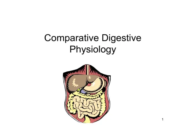
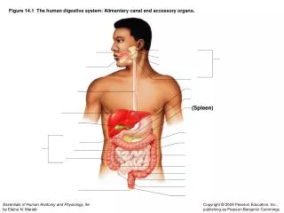
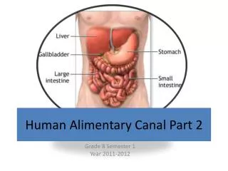
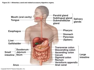
Alimentary Canal • Is the tubular portion of digestive system. • Is subdivided into: esophagus, stomach, small intestine (duodenum, jejunum and ileum), and large intestine (cecum, colon, rectum, anal canal, and appendix).
General Architectureof L/M Structure of Alimentary Canal 1- Mucosa. 2- Submucosa. 3- Muscularisexterna. 4- Adventitia OR serosa. Serosa
General Architectureof L/M Structure of Alimentary Canal or Serosa
Esophagus Four concentric layers: • Mucosa: • Epithelial Lining:Non-Keratinized Stratified Squamous Epithelium. • Lamina propria:Loose areolar C.T. with mucosal esophageal glands (secretion of mucus) in the upper and lower ends. • Muscularismucosae:Few layers of smooth muscle fibers. Serosa
Esophagus • Submucosa: • Loose areolar C.T. containing blood vessels, nerves, submucosal esophageal glands(secretion of mucus)& • Meissner’s plexus of nerve fibers and nerve cells. • MuscularisExterna: Two muscle layers: • Inner circular layer. • Outer longitudinal layer. • Upper 1/3: both layers are skeletal M. • Middle 1/3: inner layer is smooth muscle outer layer is skeletal M. • Lower 1/3: both layers are smooth M. • Auerbach’s (myenteric) plexus in between the 2 layers
Esophagus • Serosa or Adventitia: • Adventitia: is loose areolar C.T. not covered by mesothelium. • Serosa: is loose areolar C.T. covered by mesothelium (simple squamous epithelium) in the abdominal part of the esophagus. Serosa
STOMACH • It has 4 regions: cardia, fundus, body and pylorus. • Mucosa has folds, known as rugae that disappear in the distended stomach. fundus cardia body pylorus
Fundus (and Body) of Stomach • Mucosa:is invaded by fundic glands. The surface epithelium of the mucosa is simple columnar mucus-secreting cells. • Submucosa: • Connective tissue containing blood vessels, nerves, and Meissner’s plexus. • NO glands. • MuscularisExterna: • Three smooth muscle layers: • Inner oblique. • Middle circular. • Outer longitudinal. • Auerbach’s(myenteric) plexus. • Serosa: • C.T. covered by mesothelium.
Mucosa of Fundus of Stomach • It is composed of: 1. Surface Columnar Epithelium: Simple columnar epithelium: secretes mucus. 2. Lamina propria: C.T. invaded by numerous fundic glands with lymphoid elements. 3. Muscularis mucosae: 2 layers of smooth muscle fibers.
Mucosa of Fundus of Stomach Surface Columnar Epithelium
Fundic Glands • Fundic glands have: • Short pits: one fourth of mucosa. • Simple branched tubular glands. • Are rich in parietal & chief cells.
Mucosa of Fundus of Stomach 1 2 Lumen. Surface columnar epithelium. Pits of fundic glands. Fundic glands. Lamina propria. Muscularis mucosae. 3 4 5 6
Fundic Glands Composed of 5 cell types: • Parietal (oxyntic) cells. • Peptic (chief) cells. • Mucous neck cells. • Enteroendocrine(EE, DNES) cells. • Stem cells.
Fundic Glands • Parietal (oxyntic) cells: • Shape: pyramidal or polygonal. • Nucleus: central, round. • Cytoplasm: • deeply acidophilic, rich in SER and mitochondria (40% of the cell volume). • C-shaped intracellular canaliculus. • Secrete HCl and gastric intrinsic factor that helps absorption of vitamin B12. • Parietal - why? • Oxyntic - why?
Fundic Glands 2. Peptic (chief) cells: • The predominant cell type. • Columnar cells. • Nucleus: basal, round. • Cytoplasm: • basohilic with apical secretory granules. • secrete pepsinogen.
Fundic Glands 3. Mucous neck cells:secrete mucus. 4.Enteroendocrine (EE) (DNES) cells:secrete hormones (e.g. serotonin, endorphin). 5. Stem cells: regenerative cells.
Pylorus of Stomach • Mucosa: is invaded by pyloric glands. The surface epithelium is simple columnar mucus-secreting cells. • Submucosa: • Connective tissue containing blood vessels, nerves, and Meissner’s plexus. • NO glands. • MuscularisExterna: • Two smooth muscle layers: • Inner circular. • Outer longitudinal. • Auerbach’s plexus. • Serosa: • C.T. covered by mesothelium Lumen Surface epithelium Pits of pyloric glands Lamina propria Muscularis mucosae Submucosa Muscularis externa
Pyloric glands • Their pits are deep --- about half the length of mucosa. • They are branched and convoluted --- many cross sections.
Pyloric glands • Cells of pyloric glands: • Mucus neck cells (Mucus secreting cells): • The predominant cells. • Secrete mucus. • EE cells: • EC cells • G cells • D cells • A cells • Stem cells. • Parietal cells: few. • No peptic cells.