Download
1 / 89
900 likes | 905 Views
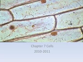
CH.7 CELL STRUCTURE AND FUNCTION. THE DISCOVERY OF THE CELL. It was not until the mid-1600 ’ s that scientists began to use microscopes to observe cells. In 1665, Englishman Robert Hooke used an early compound microscope to look at a slice of cork, plant material. CORK CELLS.

E N D
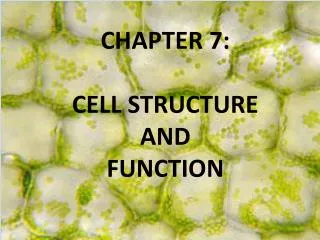
CH.7 CELL STRUCTURE AND FUNCTION
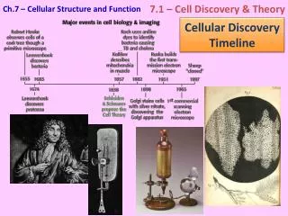
THE DISCOVERY OF THE CELL • It was not until the mid-1600’s that scientists began to use microscopes to observe cells. • In 1665, Englishman Robert Hooke used an early compound microscope to look at a slice of cork, plant material.
Hooke saw thousands of empty chambers which he called cells. They reminded him of a monastery’s tiny rooms, which were called cells. • In Holland around the same time, Antonvan Leeuwenhoek used a single-lens microscope to observe pond water and other things. • He discovered that living things seemed to be everywhere, even in the water he was drinking.
Paramecium Spirogyra
THE CELL THEORY • In 1838, German botanist MatthiasSchleiden concluded that all plants were made of cells. • In 1839, German biologist TheodorSchwann stated that all animals were made of cells.
In 1855, German physician Rudolf Virchow concluded that new cells could be produced only from the division of existing cells. • These discoveries, confirmed by other biologist, are summarized in the cell theory.
CELL THEORY STATES… 1. All living things are composed of cells. 2. Cells are the basic units of structure and function in living things. 3. New cells are produced from existing cells.
THE CHANGE OF THE MICROSCOPE • Hooke, Virchow, and others used crude microscopes to study the cells. • Today scientist can use a variety of different techniques.
High resolution video to make movies of cells as they grow. • Transmission electron microscope • electrons pass through thin slices of cell parts • Cells must be dead and in a vacuum
CONFOCAL LIGHT TEM TEM CHLOROPLAST
Scanning electron microscope • produces three-dimensional images of cells • Do not have to cut cells into slices • Vacuum TEM is more powerful than SEM
MARINE DIATOM SEM SCANNIING ELECTRON MICROSCOPE pea weevil egg
White pine sheath mite on eastern white pine Scanning electron microscope image of white pine sheath mite
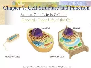
PROKARYOTES and EUKARYOTES • Cells fall into two categories, depending on whether they contain a nucleus. • Eukaryotes (YOUkaryotes)- have a nucleus. • Prokaryotes (Pro=NO)- no nucleus.
PROKARYOTIC CHARACTERISTICS • Smaller than eukaryotic cells • Genetic information is not contained by a nuclear membrane • NO internal membranes • Less complex compared to eukaryotic cells
Some glide and swim through liquids. • Forms of locomotion are: • Cilia – hair like structures • Flagella – whip like structure. ------- Example of Prokaryotes: Bacteria
EUKARYOTIC CHARACTERISTICS • Larger than prokaryotes • They usually contain dozens of structures and internal membranes = organelles • Genetic info is carried by a nucleus. • Some live solitary lives while other form large multicellular organisms. • Examples: plants, animals, fungi, and protist.
PARTS OF THE CELL • Cell biologists divide eukaryotic cell into two parts: the nucleus and the cytoplasm. • The cytoplasm is the portion of the cell that is outside the nucleus. (includes the organelles) • Cytosol = is the internal fluid of the cell, and a large part of cell metabolism occurs here
THE NUCLEUS • The nucleus is the control center of the cell. • It contains the cell’s DNA and the code for making proteins and other important molecules. • The nucleus is surrounded by a nuclear envelope composed of two membranes.
The envelope is dotted with thousands of pores, which allow materials to leave the nucleus to other parts of the cell. • Inside of the nucleus is a granular material called chromatin. • Chromatin consists of DNA bound to protein (histones).
Structure of DNA • http://www.johnkyrk.com/chromosomestructure.html
When a cell divides, the chromatin condenses to form chromosomes. • These are the distinct structures that are passed from cell to cell…generation to generation. • Deep inside the nucleus is the nucleolus which is where assembly of ribosomes begins.
? QUESTION ? • WHAT KIND OF INFORMATION IS CONTAINED IN CHROMOSOMES ? • ANSWER: Genetic Information known as DNA
RIBOSOMES • One of the most important jobs carried out in the cell is making proteins. • Proteins are assembled in ribosomes.
Ribosomes are small particles of RNA and protein found throughout the cell (mostly cytoplasm). • Ribosomes produce proteins by following coded instructions that come from the nucleus.
? QUESTION ? • WHAT DO RIBOSOMES PRODUCE ? • ANSWER: PROTEINS
ENDOPLASMIC RETICULUM • The endoplasmic reticulum also known as ER. It is the site where lipid components of the cell membrane are assembled, along with proteins and other materials.
The portion of the ER involved in synthesis of proteins is called Rough ER. • Rough ER = ribosomes found on the surface. • Newly made proteins leave ribosomes and are inserted into the Rough ER, where they can be chemically modified.