Download
1 / 38
380 likes | 546 Views
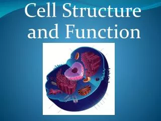
Ch. 7: Membrane Structure and Function. Introduction. The plasma membrane is selectively permeable . The macromolecules that make up the PM are lipids, proteins, and carbohydrates. Phospholipids make up most of the PM. Phospholipids are amphipathic molecules.

E N D
Ch. 7: Membrane Structure and Function
Introduction • The plasma membrane is selectively • permeable. • The macromolecules that make up the PM • are lipids, proteins, and carbohydrates. • Phospholipids make up most of the PM. • Phospholipids are amphipathic • molecules. • A model of the PM is known as the • “Fluid Mosaic Model.” • This model shows that the PM is a fluid • structure, with proteins embedded or • attached to a double layer of • phospholipids.
The PM is not rigid. There is lateral movement. • The phospholipids move rapidly. Protein movement depends on size and attachment to cytoskeleton. • Membrane fluidity also depends on temperature. Hydrophilic region of protein Phospholipid bilayer Hydrophobic region of protein
Extracellular layer Proteins Knife Plasma membrane Cytoplasmic layer Extracellular layer Cytoplasmic layer
Phospholipids with unsaturated fatty acids are more fluid. • Cholesterol maintains membrane fluidity. Lateral movement (~107 times per second) Flip-flop (~ once per month) Movement of phospholipids Viscous Fluid Saturated hydro- carbon tails Unsaturated hydrocarbon tails with kinks Membrane fluidity Cholesterol Cholesterol within the animal cell membrane
Membranes are mosaics of structure and • function • Proteins determine the function of the • PM. • There are two types of membrane protein:
Integral proteins: spans the entire • membrane. N-terminus EXTRACELLULAR SIDE C-terminus CYTOPLASMIC SIDE a Helix
B. The six major functions of membrane proteins:
Glyco- protein Attachment to the cytoskeleton and extra- cellular matrix (ECM) Cell-cell recognition Intercellular joining
Peripheral proteins: They are not • embedded into the bilayer at all. • They are loosely bounded to the PM, • often to the exposed part of an • integral protein.
C. The cytoplasmic and exterior sides of the PM differ. This difference is determined as the ER builds the PM. Vesicles fuse with the PM enlarging the PM.
Membrane carbohydrates are important • for cell-cell recognition. 1. Cells recognize other cells by keying on surface molecules, often carbohydrates, on the plasma membrane. • Membrane carbohydrates are usually • branched oligosaccharides with fewer • than 15 sugar units. • They may be covalently bonded either • to lipids, forming glycolipids, or, more • commonly, to proteins, forming • glycoproteins.
The oligosaccharides on the external • side of the plasma membrane vary from • species to species, individual to • individual, and even from cell type to • cell type within the same individual. Example: Four human blood types (A, B, AB, O) • Traffic Across Membranes • The membrane’s organization results in • selective permeability. • Sugars, amino acids, and other • nutrients enter a muscle cell and • metabolic waste products leave.
The cell absorbs oxygen and expels • carbon dioxide. • It also regulates concentrations of • inorganic ions, like Na+, K+, Ca2+, and • Cl-, by shuttling them across the • membrane. • Permeability of a molecule through a • membrane depends on its interactions • with the hydrophobic core of the membrane. • Hydrophobic molecules can easily • dissolve through the bilayer (hydro- • carbons, CO2, and O2).
2. Ions and polar molecules pass through with difficulty. Ex. Water & glucose • Proteins assist and regulate the transport of ions and polar molecules. • Polar molecules are transported across • the membrane are called transport • proteins. • Some transport proteins have hydro- • philic channels that are used as tunnels. • Each transport protein is specific as to • the substances that it will translocate • (move).
Passive transport is diffusion across a • membrane. • Diffusion is the tendency of molecules • of any substance to spread out in the • available space Molecules of dye Membrane (cross section) WATER Net diffusion Net diffusion Equilibrium Diffusion of one solute Diffusion is random, until equilibrium is reached.
A substance will diffuse down its • concentration gradient. Substance will • diffuse from where it is more • concentrated to where it is less • concentrated, down its concentration • gradient. Net diffusion Net diffusion Equilibrium Equilibrium Net diffusion Net diffusion Diffusion of two solutes
The diffusion of a substance across a • biological membrane is passive • transport because it requires no • energy from the cell to make it happen. • Osmosis is the passive transport of water. 1. A solution with the higher concentration of solutes is hypertonic. 2. A solution with the lower concentration of solutes is hypotonic. 3. Solutions with equal solute concentrations are isotonic.
Water molecules will move from a hypotonic solution to a more hypertonic solution. It will move across a selectively permeable membrane until the solutions are isotonic.
Cell survival depends on balancing water • uptake and loss. • An animal cell immersed in an isotonic • environment experiences no net • movement of water across its plasma • membrane. Water flows across the • membrane, but at the same rate in both • directions. • An animal cell in a hypertonic environ- • ment will loose water, shrivel, and • probably die. • A cell in a hypotonic solution will gain • water, swell, and burst.
Plant cells have a cell wall that contribute to its water balance. If a cell is isotonic to it surroundings, there is no movement of water into the cell and the cell is flaccid and the plant may wilt. In a hypertonic solution, a cell wall has no advantages. Plasmolysis will take place.
For a cell living in an isotonic environ- • ment (for example, many marine • invertebrates) osmosis is not a problem. • The cells of most land animals are bathed • in an extracellular fluid that is isotonic to • the cells. • Organisms without rigid walls have • osmotic problems in either a hypertonic • or hypotonic environment and must have • adaptations for osmoregulation to • maintain their internal environment. Example: Paramecium are hypertonic to their environment. • Water will continually enter the paramecium.
Paramecium have a specialized organelle, the contractile vacuole, that functions as a pump to force water out of the cell.
Specific proteins facilitate the passive • transport of water and hydrophilic solutes. • When polar molecules pass through the • membrane via proteins, this is called • facilitated diffusion. • Transport proteins are specialized for the • solute it transports. It is solute-specific. 3. There are two models of facilitated diffusion: • Channel proteins: allows for fast • transport. Ex. Aquaporins
Gated channels: a stimulus causes • them to open or close (chemical or • electrical).
Fig. 8.16 Both diffusion and facilitated diffusion are forms of passive transport of molecules down their concentration gradient, while active transport requires an investment of energy to move molecules against their concentration gradient.
Active transport is the pumping of solutes • against their concentration gradients. • Some transport proteins can move • solutes against their concentration • gradient. • Active transport requires the cell to • expend its own metabolic energy. • ATP supplies the energy for active • transport. • Active transport is performed by specific • proteins embedded in the membranes. • Active transport is a way in which cells • can maintain differing concentrations of • solutes inside the cell and outside the • cell.
Example: Sodium-Potassium pump This pump maintains higher concentrations of K+ inside the cell and lower concentrations of Na+ inside the cell. The sodium-potassium pump uses the energy of one ATP to pump three Na+ ions out and two K+ ions in.
Some ion pumps generate voltage • All cells have voltages across the • membrane. Voltage is electrical potential • energy – a separation of opposite • charges. • The inside of a cell is negative • compared to the outside of the cell. • There are more anions inside than • outside the cell. • This voltage across the membrane is called membrane potential. • The membrane potential and chemical • concentration gradient together form • an electrochemical gradient.
An ion will diffuse down its electro- • chemical gradient: • When gated channels open, Na+ ions will diffuse into the cell down the electrochemical gradient. • The sodium-potassium pump helps • maintain an electrochemical gradient • (3 Na+ pumped out for every 2 P+ • pumped into the cell). • Transport proteins that generate voltage across the membrane are called electrogenic pumps.
Another example of the electrogenic • pump is the proton pump. It actively • transports hydrogen ions (protons) out • of the cell. Proton pumps are found in mitochondria and chloroplasts. These pumps store energy that is later used for cellular work.
Cotransport: a membrane protein couples • the transport of two solutes. A substance that has been pumped across a membrane can do work; as it diffuses back, it can power another transport protein.
Exocytosis and endocytosis transport • large molecules • Large molecules like proteins and poly- • saccharides cannot be transported via • proteins. They are exported out of the • cell via exocytosis. • A transport vesicle budded from the • Golgi apparatus is moved by the cyto- • skeleton to the plasma membrane. The vesicle will fuse with the PM and • expel the contents of the vesicle. • A cell takes in macromolecules via • endocytosis.
There are three types of endocytosis: • Phagocytosis: • Pinocytosis:
Receptor-mediated endocytosis: This type of endocytosis is very specific. Special receptors bind to ligands that are in the extracellular space. Receptor proteins are usually clustered in regions of the membrane called coated pits, which are lined on their cytoplasmic side with protein.
An example of receptor-mediated endocytosis is cholesterol. Cholesterol is taken in for the synthesis of membranes and as a precursor for steroids. Cholesterol travels in blood in particles called low-density lipoproteins (LDLs). These particles bind to LDL receptors and then enter the cells by endocytosis. • Hypercholesterolemia: defective LDL receptors
Passive Transport Active Transport NO NO NO YES YES YES YES YES YES NO NO Polar – Yes Nonpolar - No