Download
1 / 17
260 likes | 1.24k Views
THE BIOLOGY OF ANAPHYLAXIS. LFSC 609E JASON RUCKER. OUTLINE. Introduction The Biology of Anaphylaxis Gell-Coombs Responses Type 1 Allergic Reactions and Anaphylaxis Biochemical Pathways Conclusion References. INTRODUCTION.

E N D
THE BIOLOGY OF ANAPHYLAXIS LFSC 609E JASON RUCKER
OUTLINE • Introduction • The Biology of Anaphylaxis • Gell-Coombs Responses • Type 1 Allergic Reactions and Anaphylaxis • Biochemical Pathways • Conclusion • References
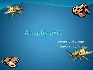
INTRODUCTION • Anaphylaxis is defined as a life-threatening allergic reaction set in action by a wide range of antigens and involving multiple organ systems. The true incidence is difficult to estimate, but in 1973 the Boston Collaborative Drug Surveillance Program reported six anaphylactic reactions and 0.87 deaths from anaphylaxis per 10,000 patients. Reactions to insect stings alone are responsible for at least 50 deaths in the United States each year.1 These figures reveal the importance of continued research into the biology of anaphylaxis along with developing new (and improving existing) therapies.
BIOLOGY OF ANAPHYLAXIS • Anaphylaxis is brought about by the release of mediators such as histamine from the mast cells and basophils. These actions are responsible for the immediate symptoms of anaphylaxis. • A late-phase response initiated by the mobilization of other inflammatory cells, including eosinophils, can occur hours after the initial reaction. • Anaphylaxis is considered an IgE-mediated allergic reaction to such things as foods, insect venoms, drugs, and latex.1 www.postgradmed.comissues199608_96wyatt1.gif
BIOLOGY OF ANAPHYLAXIS • The mast cell can be persuaded to react by additional means, including: • (1) direct activation by exercise, opiates, and possibly radiocontrast agents • (2) disturbances of arachidonic acid metabolism by cyclo-oxygenase inhibitors such as aspirin and nonsteroidal anti-inflammatory drugs (NSAIDs), and • (3) complement activation by immune complexes, caused by reactions to blood products and resulting in production of C3a and C5a.1 http://www.britannica.com/nobelprize/article-215500
BIOLOGY OF ANAPHYLAXIS • Anaphylaxic symptoms can occur within seconds of antigen exposure. With fatal reactions, the respiratory and cardiovascular systems are affected. • Upper airway obstruction caused by angioedema usually lead to asphyxia. Lower airway obstruction with wheezing and chest tightness is caused by bronchospasm. • Hypotension is caused by a shift of fluid from the intravascular to the extravascular space. • Losses of intravascular volume can occur quickly as a result of increased vascular permeability. • Patients normally compensate through maximal vasoconstriction initiated by the release of catecholamines and angiotensin.1 www.pennhealth.com...allergyimages19320.jpg
GELL-COOMBS RESPONSES • The manifestations of a particular allergic reaction depend on which of the immune mechanisms predominates in the response. Based on this criterion, immunologists use the Gell-Coombs classification system to recognize four types of hypersensitivity reactions. • Types I, II, and III involve antibody-mediated mechanisms and are of rapid onset. • The type IV reaction stems from cell-mediated mechanisms and has a delayed onset.1,2 Immunology, 5th Edition. New York. W.H. Freeman and Company
TYPE I HYPERSENSITIVITY • A Type I Hypersensitive reaction is mediated by IgE antibodies whose Fc region binds to receptors on the mast cells or blood basophils. • Crosslinkage of the fixed IgE by allergen leads to mast cell or basophil degranulation with release of pharmologically active mediators. • The primary effects of these mediators are smooth-muscle contraction and vasodilation. • Symptoms of type I reactions include potentially life- threatening systemic anaphylaxis and localized response such as hay fever and asthma.2 www-immuno.path.cam.ac.uk/.../lec13/type2a.gif
TYPE II HYPERSENSITIVITY • Type II Hypersensitive reactions occurs when antibodies reacts with antigenic determinants present on the surface of the cell. • This results in cell damage or cell death through complement-mediated lysis or antibody-dependent cell-mediated cytotoxic (ADCC).2 www-immuno.path.cam.ac.uk/.../lec13/type2a.gif
TYPE III HYPERSENSITIVITY • Type III, or immune-complex, Hypersensitive reactions are represented by tissue damage caused by the activation of complement in response to antigen-antibody (immune) complexes that are deposited in tissues. • The classes of antibody involved are the same ones that participate in type II reactions—IgG and IgM—but the mechanism by which tissue damage is brought about is different. The antigen to which the antibody binds is not attached to a cell. • Once the antigen-antibody complexes form, they are deposited in various tissues of the body, including the blood vessels, kidneys, lungs, skin, and joints. • Deposition of the immune complexes causes an inflammatory response, causing the release of tissue-damaging substances, such as enzymes that destroy tissues locally, and interleukin-1, which, among its other effects, bringing about fever.2 www-immuno.path.cam.ac.uk/.../lec13/type2a.gif
TYPE IV HYPERSENSITIVITY • A Type IV Hypersensitivity reaction involves the cell-mediated branch of the immune system. • Antigen activation of sensitized TH1 cells induces release of various cytokines that cause macrophages to accumulate and activate. • The net effect of the activation of macrophages is to release lytic enzymes that cause localized tissue damage.2 Immunology, 5th Edition. New York. W.H. Freeman and Company
BIOCHEMICAL PATHWAYS • Research in mice, which have a similar immune system to humans, had hinted that high amounts of nitric oxide (NO) throughout the body could play a role in anaphylactic attacks. • Researcher Peter Brouckaert et al induced anaphylactic shock in mice using two methods: by injecting a molecule to deliberately lower blood pressure, and by creating an allergic reaction similar to those experienced in humans. • By injecting nitric-oxide blockers into some of the mice before attempting to give them anaphylactic shock, the researchers were able to confirm that nitric oxide was the cause.3
BIOCHEMICAL PATHWAYS • Researchers believed at one time that a protein called Inducible Nitric Oxide Synthase(iNOS) was the top suspect for anaphylactic shock. • Brouckaert and his team determined that Endothelial Nitric Oxide Synthase(eNOS), a protein previously thought to produce only small amounts of nitric oxide in order to regulate the body's normal blood pressure variations, was the actual cause of anaphylactic shock. • Mice genetically engineered to lack eNOS were immune to potentially fatal allergens, whereas mice missing iNOS fell victim to them.3
BIOCHEMICAL PATHWAYS • Another biological pathway is the Complement System. It contains a group of serum proteins, many of which exists in inactive forms. • Activation occurs by the Classical, Alternative, or Lectin pathways, each of which is launched differently. • The three pathways merge in a sequence of events that lead to the production of a molecular complex that causes cell lysis.2 www.britannica.com
BIOCHEMICAL PATHWAYS • The Classical pathway is initiated by antibody binding to a target cell; reactions of IgM and certain IgG subclasses activate this pathway. • Activation of the Alternate and Lectin pathways is antibody-independent. These pathways are initiated by reaction of complement proteins with surface molecules of microorganisms.2 • Complement can opsonize bacteria for enhanced phagocytosis; it can recruit and activate various cells including polymorphonuclear cells (PMNs) and macrophages; it can participate in regulation of antibody responses and it can aid in the clearance of immune complexes and apoptotic cells. • Complement can have detrimental effects for the host because it contributes to inflammation and tissue damage and it can trigger anaphylaxis.4 Immunology, 5th Edition. New York. W.H. Freeman and Company
CONCLUSION • Anaphylaxis is a potentially fatal allergic reaction. Causes can range from bee stings to drugs, foods, and exercise. • Onset is usually sudden, and a delayed reaction can occur several hours after the first reaction. • Treatment consists of airway maintenance and support of the blood pressure with fluid expanders, epinephrine, and oxygen. • Additional medicines, such as corticosteroids, antihistamines, vasopressors, glucagon, atropine sulfate, and isoproterenol hydrochloride, may be beneficial. • Prevention is the most important part of anaphylaxis management.1 health.yahoo.commediahealthwiseh9991075.jpg
REFERENCES • Immune System Disorder http://www.britannica.com/nobelprize/article-215500. Accessed 20070804. • Goldsby. Richard T. and Thomas J. Kindt. (2003). Immunology, 5th Edition. New York. W.H. Freeman and Company. • Mice saved from lethal allergic reaction.http://www.vetscite.org/publish/items/003112/index.html. Accessed 20070804. • COMPLEMENThttp://pathmicro.med.sc.edu/ghaffar/complement.htm. Accessed 20070804.