Download
1 / 10
100 likes | 108 Views
The purpose of this study was to evaluate the Hepatoprotective activity of Punarnava Linn<br>formulations, which are well known for their hepatoprotective activity. In this study carbon<br>tetrachloride is used as a Hepatotoxicity inducer. For evaluating hepatoprotective activity<br>“carbon tetrachloride induced liver fibrosis in rats “method was selected. After the experimental<br>work the blood collected from the rats is used for determination of bio-chemical parameters like<br>SGOT-Serum Glutamate Oxaloacetate Transminase, SGPT-Serum Glutamate pyruvate<br>transaminase, ALP-Alkaline phosphatase, direct and total bilirubin. These results indicated that<br>the formulation Punarnava Linn provides significant protection against the toxic effect of carbon<br>tetrachloride on liver. The reduction in the elevated enzyme levels suggests the protection of<br>liver cells by Punarnava Linn drugs.<br>

E N D
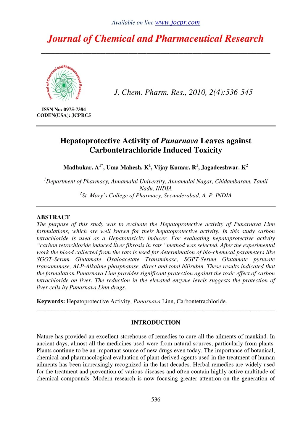
Available on line www.jocpr.com Journal of Chemical and Pharmaceutical Research __________________________________________________ J. Chem. Pharm. Res., 2010, 2(4):536-545 ISSN No: 0975-7384 CODEN(USA): JCPRC5 Hepatoprotective Activity of Punarnava Leaves against Carbontetrachloride Induced Toxicity Madhukar. A1*, Uma Mahesh. K1, Vijay Kumar. R1, Jagadeeshwar. K2 1Department of Pharmacy, Annamalai University, Annamalai Nagar, Chidambaram, Tamil Nadu, INDIA 2St. Mary’s College of Pharmacy, Secunderabad, A. P. INDIA ABSTRACT The purpose of this study was to evaluate the Hepatoprotective activity of Punarnava Linn formulations, which are well known for their hepatoprotective activity. In this study carbon tetrachloride is used as a Hepatotoxicity inducer. For evaluating hepatoprotective activity “carbon tetrachloride induced liver fibrosis in rats “method was selected. After the experimental work the blood collected from the rats is used for determination of bio-chemical parameters like SGOT-Serum Glutamate Oxaloacetate Transminase, SGPT-Serum Glutamate pyruvate transaminase, ALP-Alkaline phosphatase, direct and total bilirubin. These results indicated that the formulation Punarnava Linn provides significant protection against the toxic effect of carbon tetrachloride on liver. The reduction in the elevated enzyme levels suggests the protection of liver cells by Punarnava Linn drugs. Keywords: Hepatoprotective Activity, Punarnava Linn, Carbontetrachloride. ______________________________________________________________________________ INTRODUCTION Nature has provided an excellent storehouse of remedies to cure all the ailments of mankind. In ancient days, almost all the medicines used were from natural sources, particularly from plants. Plants continue to be an important source of new drugs even today. The importance of botanical, chemical and pharmacological evaluation of plant-derived agents used in the treatment of human ailments has been increasingly recognized in the last decades. Herbal remedies are widely used for the treatment and prevention of various diseases and often contain highly active multitude of chemical compounds. Modern research is now focusing greater attention on the generation of 536
Madhukar. Aet al J. Chem. Pharm. Res., 2010, 2(4):536-545 _____________________________________________________________________________ scientific validation of herbal drugs based on their folklore claim. In this modern era, a large Indian population still relies on the traditional system of medicine, which is mostly plant based. Free radical initiating auto oxidation of cellular membrane lipids can lead to cellular necrosis and is now accepted to be important in connection with a variety of pathological conditions [1]. Liver is an aerobic organ which generates reactive oxygen species that induce oxidative tissue damage. These radicals react with cell membranes and induce lipid per oxidation or cause inflammation, which may result as important pathological mediators in many clinical disorders such as heart disease, diabetes, gout and cancer [2]. Reduction of these radicals by antioxidant molecules is crucial to the protection of cells against various disorders. Development of life threatening diseases like cancer and also liver disorders are linked to the availability of these antioxidants. Flavonoids are phenolic compounds widely distributed in plants, and have been reported to exert multiple biological effects, including antioxidant and free radical scavenging abilities [3]. In spite of tremendous advances in modern medicine no effective drugs are available, which stimulate liver functions and offer protection to the liver from the damage or help to regenerate hepatic cells [4]. In absence of reliable liver-protective drugs in modern medicine, a large number of medicinal preparations are recommended for the treatment of liver disorders [5] and quite often claimed to offer significant relief. Attempts are being made globally to get scientific evidences for these traditionally reported herbal drugs. Punarnava Linn (Nyctaginaceae), otherwise known as “Rakta Punarnava, hogweed, boerhoavia dissusa, B.erecta” in Ayurvedic system of medicine. It is found wild throughout India and srilanka, Punarnava Linn is found in Himalayan valleys up to 2000-2500m. The weed is also grown in Malaysia, china and Africa. Leaves are 25-30 mm long and 12-20 mm in wide, ovate, oblong sub-orbicular, and apex slightly pointed with rounded or sub-cordate base. Leaves are glabrous, rather thick in texture and petiolate. Petioles are double the length of the blade. Margin is entire [6]. The present study aims in the evaluation of hepatoprotective and in-vitro cytotoxic activity of alcoholic extract of leaves of Punarnava Linn. EXPERIMENTAL SECTION Following is the list of natural products used by Ayurvedic physicians for their hepatoprotective activity. 2.1. Plant materials The leaves of Punarnava Linn (Nyctaginaceae)Linn were collected in the month of October - December. The leaves were shade dried, coarsely powdered and subjected to successive extraction using n-hexane and alcohol in soxhlet apparatus. Both the extracts were then dried under reduced pressure and the percentage yields of the extracts were 8.2%, 20% respectively. Hepatoprotective and in-vitro cytotoxic activity were carried out by using alcoholic extract. 2.2. Experimental animals 30 male Albino rats of Wister strain weighing 150-200 g were used for the study. They were maintained under standard environmental condition (temperature 25-280C and 12h light/dark cycle) and they were allowed with standard laboratory feed and water ad libitum. The animals were given a week’s time to get acclimatized with the laboratory condition. Initial body weights of all animals were recorded. Ethical clearance for the use of animals was obtained from the committee constituted for the purpose. 537
Madhukar. Aet al J. Chem. Pharm. Res., 2010, 2(4):536-545 _____________________________________________________________________________ 2.3. Hepatoprotective activity A total of 30 Albino rats were divided into 5 groups of 6 animals each: Group A: Vehicle control andall other groups were given Carbontetrachloride (1 ml/kg) for two successive days (i.e. 2 & 3 days of 7 day’s course) Group B: Receives only Carbontetrachloride Group C: (Carbontetrachloride 1 ml/kg + Arq biranjasif3ml/kg) Group D: (Carbontetrachloride 1 ml/kg + Lohasav 1 ml/kg and Punarnava 1 ml/kg. Group E: (Carbontetrachloride 1ml/kg + Dabadulward 500 mg/kg) drug are administered for seven days according to the body weight After drug treatment the animals are sacrificed, blood is collected. Blood serum is used for estimation of biochemical parameters. 2.4. Different methods available for evaluating hepatoprotective activity are: i). Allyl alcohol Induced Liver fibrosis Procedure: Female Wister rats weighing 120-150gms are used. At 8 am of the first day food but not water is withdrawn. At 3.00pm the compounds to be tested for protective activity are administered i.p or orally. One hour later, the animals are dosed orally with 0.4ml/kg of 1.25% solution of ally alcohol in water. At 80 am of the second day treatment with the potentially protective drugs is repeated. Food but not water is withheld until the third day. At 8am of the third day the animal are sacrificed and the liver is removed. The parietal sides of the liver (left, medium and right lobe caudatus) are checked using stereomicroscope with 25 times magnification. Focal necrosis is observed as white-green or yellowish hemorrhagic areas clearly separated from unaffected tissue. The diameter of necrotic areas is determined using a ocular micrometer. These values are added for each animal to obtain an index for necrosis. ii). Bile duct ligation induced liver fibrosis Procedure: Male Sprague Dawley rats weighing approximately 250 Gms with ketanest (rompun). Laparatomy is performed under antiseptic conditions. A midline incision in the abdomen is made from the xiphostermum to the pubis, exposing the muscle layers and the linea alba, which is then incised over a length corresponding to the skin incision. The edges of the liver are then raised and the duodenum pulled down to expose the common bile duct which pursues an almost straight course of about 3 cm from the hilum of the liver to its opening into the duodenum. There is no gallbladder, and the duct is embedded for the greater part of its length in the pancreas, which opens into it by numerous small ducts. A blunt aneurysm needle is passed under the part of the duct selected; stripping the pancreas away with care, and duct is divided between double ligatures of cotton thread. The peritoneum and the muscle layers as well as skin mould are closed with cotton stitches. The animals receive normal diet and water throughout the experiment. Groups of 5-10 animals receive the test compound in various doses or the vehicle twice daily for 5 weeks then, they are sacrificed and blood is harvested for determination of bile acids, 7s fragment of type iv collagen, and procollagenin-peptide. The liver is used for histological examination and for hydroxyproline determinations. Control animals show excessive bile duct proliferation as well as formation of fibrous septa. The picture is complicated is consistent with complete biliary cirrhosis. iii) Galactosamine induced Liver fibrosis Procedure: For induction of acute experimental hepatotoxicity, divided doses of 100-400mg/kg D-galactosamine are injected to rats' i.p. or i.v. during one day. For induction of liver cirrhosis, male Wister rats weighing 110-180 gm are injected intraperitoneally 3 times weekly with 500 mg/kg D-galactosamine over a period of 1-3 months. Potential protective substances are 538
Madhukar. Aet al J. Chem. Pharm. Res., 2010, 2(4):536-545 _____________________________________________________________________________ administered orally with the food or by gavage every day. The rats are sacrificed at various time intervals & the livers obtained by autopsy. iv). Carbon tetrachloride Induced liver fibrosis in Rats This method was followed in the present study. Assessment of hepatoprotective activity is carried out in Wister rats. The animals were segregated into five groups of six animals each. Group A served as normal control. All other groups received Carbonterachloride (1ml/kg) for two successive days. Group B animals were maintained as Carbon tetrachloride group, Group C served as normal control. All other groups received Carbon tetrachloride (1ml/kg) for two successive days. Group B animals were maintained as Carbon tetrachloride group, Group D and V animals were treated with drugs for 7 days. After drug treatment all the animals were sacrificed, blood was collected by puncturing the retroarbital p0lexus and was allowed to clot for 45 min at room temperature, serum was collected by centrifugation at 2500 rpm for 15 min, used for 2.5. Estimation of various bio-chemical parameters Assessment of Liver function Bio-chemical parameters such as glutamate oxaloacetate transaminase (GOT), glutamate pyruvate transminase (GPT), alkaline phosphatase (ALP) acid phosphatase (ACP) and serum Bilirubin were determined. Livers were excised quickly fixed in 10% buffered neutral formalin and then fixed in bovine solution, they were processed for paraffin embedding following the standard micro technique. Sections of liver stained with alum-haematoxylin and eosin, were observed microscopically for histopathological changes. In the present study Carbontetrachloride Induced liver fibrosis in Rats was flowed. 30 Healthy rats of either sex weighing 150-175 g were housed in groups of six per cage a temperature of 25 ±1°C and relative humidity of 20% A12:12 hour of dark: light cycle was followed during the experiment. The experiment was carried out during 12:00 – 14.00 hours. Animals had free access to food and water, however food but not water was withdrawn while administering the drug. The rats were divided into five groups each containing six rats. 2.6. Methods for determination biochemical parameters i). Determination of Serum Glutamate oxaloacetate Transaminase (SGOT) Method: Reitman and Frankel SGOT catalyses the following reaction: α-Ketoglutarate + L-Aspartate <…….............> L- Glutamate + Oxaloacetate Oxaloacetate so formed is coupled with 2, 4-Dinitrophenyl hydrazine to give corresponding hydrazone, which gives brown colour in alkaline medium and this, can be measured colorimetrically. Reagents used: Buffered Aspartate α-Ketoglutarate substrate, pH 7.4, Dinitrophenyl hydrazine (DNPH) colour reagent, Sodium Hydroxide, Working Pyruvate standard. 539
Madhukar. Aet al J. Chem. Pharm. Res., 2010, 2(4):536-545 _____________________________________________________________________________ Table. 1: For test Procedure Pipette Into tube marked Buffered Aspartate α-Ketoglutarate Substrate Serum Mix well incubate at 3 C for 60 min Dinitropheyl hydrazine Mix well and allow to stand at Room Temperature for 20 min Dilute 1ml of NaOH to 10ml with purified water Test (T) 0.25ml 0.05ml - 0.25ml - 2.5ml Mix well and allow standing at Room Temperature for 10min and reading the optical Density against purified water on a colorimetry at 505nm. Calculation: Mark the optical density on the Y-axis of the standard curve and extrapolate it to the corresponding enzyme activity on X-axis ii). Determination of Serum Glutamate Pyruvate Transaminase (SGPT) Method: Reitman and Frankel SGPT (ALT) catalyses the following reaction: α-Ketoglutarate + L-Alanine <……………> L- Glutamate + Pyruvate Pyruvate so formed is coupled with 2, 4-Dinitrophenyl hydrazine (2, 4 DNPH) to give corresponding hydrazone, which gives brown colour in alkaline medium and this can be measured colorimetrically. Reagents used: Buffered Alanine α-Ketoglutarate Substrate, pH 7.4, Dinitrophenyl hydrazine (DNPH) colour reagent, Sodium Hydroxide, 4N, Working Pyruvate Standard 2m M Table. 2: For Test Procedure Pipette Into tube marked Buffered Aspartate α-Ketoglutarate Substrate Serum Mix well incubate at 3 C for 60 min Dinitropheyl hydrazine Mix well and allow to stand at Room Temperature for 20 min Dilute 1ml of NaOH to 10ml with purified water Mix well and allow standing at Room Temperature for 10 min and reading the optical Density. Against purified water on a colorimetry at 505 nm. Calculation: Mark the optical density on the Y-axis of the standard curve and extrapolate it to the corresponding enzyme activity on X-axis. iii). For the Determination of Direct &Total Bilirubin in Serum Method: Mod .Jendrassik and Grof’s method Test (T) 0.25ml 0.05ml - 0.25ml - 2.5ml 540
Madhukar. Aet al J. Chem. Pharm. Res., 2010, 2(4):536-545 _____________________________________________________________________________ Bilirubin is mainly formed form the Heame portion of aged or damaged RBC’s. It then combines with albumin to form a complex which is not water soluble. This is refereed to as unconjugated Bilirubin. In the liver this Bilirubin complex is combined with glucuronic acid into a water- soluble conjugate. This is referred to as conjugated Bilirubin. Elevated levels of Bilirubin are found in liver diseases (Hepatitis, cirrhosis), excessive hemolysis / destruction of RBC (hemolytic jaundice) obstruction of the biliary tract (obstructive jaundice) and in drug induced reactions. The differentiation between the direct and indirect Bilirubin is important in diagnosing the cause of hyperbilirubionemia. Bilirubin _ Diazotized sulphanilic acid --------------------> Azobilirubin compounds. Assay Procedure: Pipette into clean dry test tubes labeled as Blank (B), and Tests (T) Table. 3 Addition Sequence Direct Bilirubin Reagent (L1) Direct Nitrite Reagent (L2) Sample Mix all and incubate at Room Temperature for exactly 5 min. Measure the absorbance of the Test samples against their respective blanks. Calculation: Total or Direct Bilirubin Assay = Absorbance. T x 13 iv). Alkaline phosphatase Method: Mod king and Kings Method Alkaline phosphatase (ALP) is an enzyme of hydrolyses class and acts in alkaline medium. It is found in high concentrations in the liver, biliary tract, epithelium and in the bones. Normal levels are age dependent and increases during level development. Increased levels are associated mainly with liver and bone disease moderate increases are seen in Hodgkin's disease and congestive heart failure. Table. 4 Addition Sequence B(ml) Distilled water 1.05 Buffer Reagent 1.0 Substrate reagent 0.10 Mix well and allow to stand at 37°C for 3 minutes and add Sample - Phenol reagent - Mix well and allow to stand at 37°C for 15 minutes and add Colour reagent 1.0 Sample - B (ml) 1.0 - 0.1 T(ml) 1.0 0.05 0.1 S(ml) 1.00 1.0 0.10 - C (ml) 1.0 1.0 0.10 - T (ml) 1.0 1.0 0.10 - - - - - - 0.05 - - 0.05 - - 1.0 - 1.0 0.05 1.0 - 541
Madhukar. Aet al J. Chem. Pharm. Res., 2010, 2(4):536-545 _____________________________________________________________________________ Di sodium phenylphosphatase + H2O ALP, pH 10 Phenol + di sodium hydrogen phosphate. Phenol + 4-Aminoantipyrine alkaline medium red coloured complex potassium ferricyanide. Assay Procedure: Pipette into four clean dry test tubes labeled as blank (B) standard (S) control (C), Test (T) table. Mix well after each addition measures the absorbance of the blank (Abs. B), standard (Abs, S), control (Abs.C) and test (Abs.T) against distilled water. Calculation: Abs. T-Abs.C Total ALP activity in K. A units = –––––––––––––– Abs. S-Abs.B RESULTS AND DISCUSSION Results indicated that the formulation Punarnava provides significant protection against the toxic effect of CCl4 on liver. In CCl4 induced toxic hepatitis, toxicity begins with the changes in endoplasmic reticulum, which results in the loss of metabolic enzymes located in the intracellular structures. The toxic metabolite CCl3 radical produced by microsomal oxidase system, binds covalently to macromolecule and causes per oxidative degradation of lipid membranes of the adipose tissues. The blood samples of the group B animal showed drastic increase of Serum Glutamate Oxaloacetate Transaminase (134.8±3.83), Serum Glutamate Pyruvate Transaminase (133.5 ± 7.66), Alkaline phosphatase (348.8 ± 0.74), Direct Bilirubin (0.236 ± 0.022), Total Bilirubin (0.80 ±0.016) Accumulation of higher concentrations of Bilirubin confirms the depth of, and intensity of the liver damage. Administration of Punarnava showed recovery against the toxic effects of CCl4 as shown in the table 4.6.SGOT levels reduced to 81.33±0.89, 77.83±0.965, 80.8±0.613; SGPT levels reduced to 78.66±1.79, 78.16±0.99, 75.33±1.57; ALP levels reduced to 228.16±0.99, 204±3.33, 240.5±0.77; Total Bilirubin levels reduced to 0.76±0.007, 0.76±0.0083, 0.76±0.0028 and Direct Bilirubin levels reduced to 0.215±0.003, 0.21±0.007, 0.22±0.005 with respective to the drugs given above. Hepatocellular necrosis leads to very high level of Serum Glutamate Oxaloacetate Transaminase, Serum Glutamate Pyruvate Transaminase released from liver in to the blood. Among the two, GPT is a better index off liver injury, as liver GPT activity represents 90% of total enzyme present in the body. ALP activities on the other hand are related to the functioning of hepatocytes, Increase n its activity is due to increased synthesis in presence of increased biliary pressure. Reduction in the levels of SGOT, SGPT towards the respective normal value is an indication of stabilization of plasma membrane as well as repair of hepatic tissue damage caused by carbon tetrachloride. This effect is an agreement with view that serum levels of transaminases return to normal with healing of hepatic parenchyma and regeneration of hepatocytes. Suppression of increased ALP activity with concurrent depletion of raised Bilirubin level suggests the stability for the biliary function in rat liver during chronic hepatic injury with CCl4. Carbon tetrachloride induced Hepatotoxicity is protected and reduced with the simultaneous administration of herbal preparation Punarnava is indicated by the levels of SGPT, SGOT, ALP, Bilirubin Total and Bilirubin Direct with reference to tables. 5 and Fig. 1, 2, 3, 4, 5. This reduction in the elevated enzyme levels suggests that protection of structural integrity of Hepatocyte cell membrane by Punarnava. The effectiveness of the normal functional condition 542
Madhukar. Aet al J. Chem. Pharm. Res., 2010, 2(4):536-545 _____________________________________________________________________________ of the liver is indicated by the decreased level of Serum Bilirubin. Our results suggest that the formulations Punarnava protect the liver from severe damage caused by carbon tetrachloride. Table. 5: Comparative protective Effect of Punarnava, on Carbontetrachloride induced hepatotoxicity in Rats Group Dose SGPT (IU/L) SGOT (IU/L) ALP (IU/L) Total Bilirubin (mg. dl) 0.765± 0.025 0.80± 0.29* 0.766± 0.04 Direct Bilirubin (mg. dl) 0.206± 0.0045 0.236± 0.0061 0.213± 0.007 163± 1.75 349± 2.39# 204± 4.30# Control (Group-A) 1ml/kg 79± 2.28 135± 1.49# 78.2± 1.58# 47± 1.29 134± 1.88# 78± 1.50# Carbontetrachloride (Group-B) Carbontetrachloride +Arq biranjasif (Group-C) Carbontetrachloride +Lohasav and Punarnava(Group-D) Carbontetrachloride +Dabadulward (Group-E) Values are expressed as mean ± SEM of 6 animals in each group. *p<0.01, #p<0.001 as compared to group-A 1ml/kg 1ml/kg+ 3ml/kg 228± 1.55 0.215± 0.003 1ml/kg+ 1ml/kg, 1ml/kg 1ml/kg+ 500mg/k g 79± 1.71# 81± 1.78 0.768± 0.02 241± 2.15 0.22± 0.005 77± 1.62 81± 1.80 0.768± 0.02 SGPT (IU/L) 140 120 100 80 IU/L 60 Series1 40 20 0 GROUP A GROUP B GROUP C GROUP D GROUP E GROUPS Fig. 1: Effect of Serum Glutamate Pyruvate Transaminase (SGPT) on all Groups SGOT (IU/L) 140 120 100 80 IU/L 60 Series1 40 20 0 GROUP A GROUP B GROUP C GROUP D GROUP E GROUPS Fig. 2: Effect of Serum Glutamate oxaloacetate Transaminase (SGOT) on all Groups 543
Madhukar. Aet al J. Chem. Pharm. Res., 2010, 2(4):536-545 _____________________________________________________________________________ Alkaline phosphatase 350 300 250 200 IU/L 150 Series1 100 50 0 GROUP A GROUP B GROUP C GROUP D GROUP E GROUPS Fig. 3: Effect of Alkaline Phosphatase in Serum (ALP) on all Groups TOTAL BILIRUBIN 0.8 0.79 0.78 mg.dl 0.77 Series1 0.76 0.75 0.74 GROUP A GROUP B GROUP C GROUP D GROUP E GROUPS Fig. 4: Effect of Total Bilirubin in Serum on all Groups DIRECT BILIRUBIN 0.24 0.235 0.23 0.225 0.22 mg.dl 0.215 0.21 Series1 0.205 0.2 0.195 0.19 GROUP A GROUP B GROUP C GROUP D GROUP E GROUPS Fig. 5: Effect of Direct Bilirubin in Serum on all Groups CONCLUSION The present investigation revealed that the alcoholic extract of leaves of Punarnava Linn (Nyctaginaceae) exerted significant protection against CCl4 induced hepato toxicity by its ability to ameliorate the lipid per oxidation. The extract also showed good in-vitro cytotoxic activity. Our result indicated that the potent hepatoprotective and cytotoxic activity of the extract may be due to its antioxidant and free radical scavenging properties which may be due to presence of flavanoids in the plant. Further investigation is under progress to evaluate the in vivo anticancer activity and to determine the exact phytoconstituents that is responsible for its hepatoprotective and cytotoxic activity. REFERENCES [1]W. Pyor, Ann. Rev. Physiol. 1986; 48: 657. doi:10.1146/annurev. ph.48. 030186. 003301. [2]M. Vuillaum, Mutat. Res. 1987; 43: 186. [3]N. L. Baek, J. S. Kimys, K. L. Kyung, and K. I. Park, Korean J. Pharmacog. 1996; 27: 113. [4]Chattopadhyay R.R. J. Ethnopharmacol. 2003; 89: 217. 544
Madhukar. Aet al J. Chem. Pharm. Res., 2010, 2(4):536-545 _____________________________________________________________________________ [5]Chatterjee T.K. Medicinal Plants with Hepatoprotective Properties in Herbal Options. Books and Allied (P) Ltd. Calcutta. 2000; 3: 135. [6]S.B.Gokale, C.K.Kokate, A.P.Purolit. A Text Book of Pharmacognosy. 29th edition. 2009; 103-105. 545