Download
1 / 58
740 likes | 1.15k Views
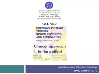
CLINICAL APPROACH TO THE DRUG-INDUCED LUNG TOXICITY. Demet KARNAK, MD Ankara University Sc h o o l of Medicine Department of Chest Disease Interstitial Lung Dis e ase Subdivision. DRUG-INDUCED LUNG TOXICITY. First mentioned in 1972 , Rosenow EC 19 drug s, pulmonary toxicity

E N D
CLINICAL APPROACH TO THE DRUG-INDUCED LUNG TOXICITY Demet KARNAK, MD Ankara University School of Medicine Department of Chest Disease Interstitial Lung Disease Subdivision
DRUG-INDUCED LUNG TOXICITY • First mentioned in1972,Rosenow EC 19 drugs, pulmonary toxicity • > 350 drugs • First runner of iatrogenic injury • Various types of injury • Mortality and morbidity↑ • Rosenow EC: Dis Mon 40:253-310, 1972 • Aronchick JM, Gefter WB: Semin Roentgenol 30:18-34, 1995
DRUG-INDUCED LUNG TOXICITY • 5% of side effects • 0,03% of hospital deaths • Discontinuation (toxicity and death↓) • Past clinical and drug history in detail • Knowledge about the drugs on the market
DRUG-INDUCED LUNG TOXICITY • Airway involvement (larynx edema, cough, bronchospasm) • Parenchymal involvement (ILD) • Pulmonary circulation (PHT, permeability ↑, vasculitis) • Pleural involvement (effusion, thickening) • Lupus syndrome • Neuromuscular disease(respiratory insufficiency) • Lymphadenopathy
RISK FACTORS • Genetic predisposition • Basic pulmonary functions • Underlying disease • Environmental factors • Activation and detoxification mechanisms • Therapy interactions and other drug toxicity • Dose
DIAGNOSTIC CRITERIA • History of drug exposure • Appropriate clinical picture • Exclusion of other diagnoses • Improvement after therapy cessation • Recurrence after re-administration
TYPES OF INJURY • Interstitial • Alveolar (diffuse lung injury, alveolar hemorrhage, edema) • Vascular
MECHANISMS OF INJURY • Oxidative • Direct toxicity • Phospholipid accumulation induced • Immune system induced
TYPES OF INVOLVEMENT • Asthma • BOOP • Hypersensitivity infiltrates • Interstitial pneumonitis-fibrosis • Non-cardiogenic pulmonary edema • Pleural effusion • Eosinophilia and pulmonary infiltrates • Pulmonary vascular disease
DRUG INDUCED LUPUS (DIL) • Underlying lupus may become manifest • Activation can occur if predisposition exists • Drug can cause lupus by itself • Fever, myalgia, rash, arthralgia, arthritis, serositis • Parenchymal and pleural involvement, 50% and 75% respectively • Pleural effusion, pleurodynia, diffuse interstitial pneumonitis, alveolar infiltrates • Should be distinguished from spontaneous lupus (SL) (F/M:1 in DIL, F/M:9/1in SL)
DRUG INDUCED LUPUS(DIL) • Discoid lesion or CNS involvement can not be seen, malar rash 2%, renal involvement 5% • Complements ve immune complexes DIL-Normal (vs. SL-↑) • antihistone ab (+) in 95% of cases but other ab (anti-ds DNA) (-) • In 90% of cases, responsible drugs are diphenilhydantoin, hydralazine, procainamide, isoniazid
Sulphonamide Hydralazine İsoniazid Procainamide Amiodarone ACEİ Beta-blocking agents Carbamazepine Gold Simvastatin Etanercept Phenytoin Tetracycline SYSTEMIC LUPUS ERYTHEMATOSUS AND LUPUS-LIKE SYNDROME
DRUG INDUCED BRONCHOSPASM • Propranolol or other beta-adrenergic antagonistsCan increase airway resistance in normal or asemptomatic asthma cases, should not be used in COPD or asthma • Thymolol Can increase airway resistance in asthmatic glocoma cases • Aspirin Causes bronchospasm in 4% of asthma cases • Other non-steroid anti-inflammatory agents Can cause similar symptoms
AMIODARONE • Primary drug causing pulmonary toxicity • Suppresses ventricular and/or supraventricular beats • ILD, OP, ARDS, solitary pulmonary nodule • High concentration oxygen along with amiodarone may lead to ARDS • Immunological mechanisms, direct toxicity or free radicals • Interstitial pneumonitis - 400mg/d • ILD - Incidental beginning, dry cough, dyspnea, weight loss and widespread patchy infiltrates
AMIODARONE • İnfiltrates are not pathognomonic • FOB and BAL excludes infections • Foamy alveolar macrophages, type 2 cell hyperplasia and fibrosis • These features can also be seen in patients without toxicity
AMIODARONE • OP- Dry cough, pleuritic type pain, fever, dyspnea, patchy infiltrates 25% • Pneumonitis, CHF, PTE should be excluded • No specific test • Galium scintigraphy is positive
ACE INHIBITORS • 3-20 % of cases • Persistant, dry cough, increasing cough at night • A couple of months or a year later (after first dose?) • Symptoms decrease in 3 days and disappear in 10 days after cessation • Pathogenesis is not clear - kinins?, substance P? • Arachidonic acid pathway activation by ACE inhibition • Thromboxan A2 elevation and bronchoconstruction • Thromboxan inhibition - stop cough
ASPIRIN • Samter syndrome (Aspirin triad) Asthma, nasal poliposis, drug sensitivity - 4% of asthma cases • Vasomotor rhinitis- clear rhinorrhea • Nasal polyps and asthma symptoms in middle ages • Aspirin sensitivity 3rd – 4th decades • Wheezing, flushing, angioedema, gastrointestinal symptoms - 30 min or 2 hrs later
ASPIRIN • Aspirin blocks cyclo-oxigenase 1 pathway which forms thromboxanes and prostoglandins from arachidonic acid. • Alternate way activates by this inhibition. • 5-lipo-oxigenase enzyme converts arachidonic acid to potent bronchoconstrictor compounds (LTC4, LTD4, LTE4)
ANALGESIC AND ANTI-INFLAMMATORY DRUGS: ASPIRIN • High doses (>30-40mg/dl, uncertain) can cause acute pulmonary edema; smoking is a risk factor? • Dyspnea, tachypnea, letargy and confusion, hypoxemia • Obvious cardiomegaly or perihilar alveolar infiltration without pleural fluid • Increased alveolocapillary permeability • 1-2% mortality in youngs, 25% in elder cases
TRANSFUSION LUNG INJURY (TRALI) • Develops a few hours later from tx • Donor originated ab can cause the disease • These antibodies are HLA (Class I or II) or granulocyte-specific • These antibodies appear to act as mediators, which result in granulocyte aggregation, activation, and microvascular pulmonary injury • In a retrospective study, these antibodies were detected in 85% of donors • 80% of patients recover within 96 hours of the original insult • No permanent pulmonary sequelae • Diagnosis can be established by detecting donor originated ab in donor circulation Kopko PM, et al: JAMA 287:1968-71, 2002
ALVEOLAR HEMORRHAGE • After diffuse hemorrhage in pulmonary capillary bed, rare • Incidental BAL diagnosis, subclinic pulmonary infiltrates, hemociderin laden macrophages, ARDS • Unexplained anemia or transient hypoxemia attacks
MICROSCOPIC POLYANGITIS • Anti-thyroid drugs - PTU and thiamazol (methimazol) • Mimics Wegener granulamatosis • Antimyeloperoxidase ab (p-ANCA) positivity • After discontinuation, clinical and immunological symptoms improve
Alveolar hemoraji • Interstitial and alveolar infiltrates causing dyspnea in advanced cases • Increased DLCO, anemia • Hemorrhage or hemociderin laden macrophages in BAL • Otoimmune disease can be excluded by detecting otoantibodies
LEUKOTRIENE ANTAGONISTS • Steroid cessation and LTA administration can lead to Churg-Strauss syndrome in asthma cases • Pulmonary infiltrates, cardiomyopathy and eosinophilia can relieve after discontinuation of LTA and commencing steroids Limper AH, Rosenow EC.In Murray JF,Nadel JA. Textbook of Respiratory Medicine, 2000 1971-94
PULMONARY VENO-OCCLUSIVE DISEASE • Iatrogenic disease is extremely rare • Bleomycin, carmustine, gemcytabine, mitomycin, vinca alcoloids, radiation and bone marrow tx • Dyspnea, irregular interstitial changes, Kerley-B lines • PHT and right heart failure
L-TRYPTOPHAN AND EOSINOPHILIA MYALGIA SYNDROME • High doses of L-tryptophan causes EMS • Insidious/rapid onset myalgia, skin changes, and cardiac-neurologic constitutional symptoms can occur • Pulmonary infiltrates, PHT, pleural effusion and acute respiratory insufficiency • Histology; eosinophilic infiltration and angitis • Symptoms relieve by cessation or sometimes be permanent • May be mortal
CHEMOTHERAPY RELATED LUNG INJURY • Generaly 2 months after discontinuation . • Main picture - “pulmonary fibrosis” Lacework honey-combing Irregular septal thickening Intralobular lining Marked bronchioles Raugh honey-combing Alteration in anatomy Fibrosis
VINCA ALCALOIDS • Combination therapy can cause the injury • 387 cases; 25 cases developed acute dyspnea and 87% of cases had focal interstitial infiltrates and reduced DLCO • 60% of cases have chronic respiratory insufficiency • Bronchospasm • Progressive interstitial fibrosis following radiation Luedke D, et al: Cancer 55:542-545, 1985 Rivera MP, et al: Am J Clin Oncol 18:245-250, 1995 Davies AM, et al: Invest New Drugs Apr 18; 2007 (in press)
Alkylating drugs • Low molecular weight compounds produce direct toxic effect when they pass through pulmonary circulation and they cause interstitial pneumonitis and consequently fibrosis • Bisulphan • Cyclophosphamide • Melphalan • Chlorambusil • BCNU (carmustine)
Bleomycin • Binds Fe+2 and forms Fe+3 • Fe+3 + O2 complex produces free radicals • PG + LTx activation • IL-1, MIP-1, MCP-1, TGF-b, TNF-a areincreased • Redox reaction and fatty acid oxidation • Membrane instabilization • Capillary endothelium damage and apoptosis • Interstitial edema • Type 1 pneumocyte necrosis • Type 2 pneumocyte metaplasia and fibrosis • Bleomycin hydrolase • Liver, spleen, bone marrow, GİS (+) • Lung and skin (-)