Download
1 / 37
390 likes | 587 Views
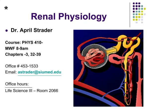
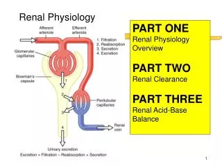
Renal Physiology Dr. Eman El Eter. The Urinary System. Functions of the urinary system Anatomy of the kidney Urine formation glomerular filtration tubular reabsorption water conservation Urine and renal function tests. Urinary System. Two kidneys. Two ureters. Urethra.

E N D
The Urinary System • Functions of the urinary system • Anatomy of the kidney • Urine formation • glomerular filtration • tubular reabsorption • water conservation • Urine and renal function tests
Urinary System • Two kidneys • Two ureters • Urethra
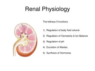
Kidney Functions • Filters blood plasma, eliminates waste, returns useful chemicals to blood • Regulates blood volume and pressure • Regulates osmolarity of body fluids • Secretes renin, activates angiotensin, aldosterone • controls BP, electrolyte balance • Secretes erythropoietin, controls RBC count. • Formation of the active form of vitamin D • Regulates acid base balance • Detoxifies free radicals and drugs • Gluconeogenesis
Nitrogenous Wastes • Urea • proteinsamino acids NH2 removed forms ammonia, liver converts to urea • Uric acid • nucleic acid catabolism • Creatinine • creatinine phosphate catabolism • Renal failure • azotemia: nitrogenous wastes in blood • uremia: toxic effects as wastes accumulate
Excretion • Separation of wastes from body fluids and eliminating them • respiratory system: CO2 • integumentary system: water, salts, lactic acid, urea • digestive system: water, salts, CO2, lipids, bile pigments, cholesterol • urinary system: many metabolic wastes, toxins, drugs, hormones, salts, H+ and water
Anatomy of Kidney • Renal cortex: outer 1 cm • Renal medulla: renal columns, pyramids - papilla • Lobe of kidney: pyramid and it’s overlying cortex
Nephrons • Types of nephrons: • Cortical nephrons (85%) • short nephron loops • efferent arterioles branch off peritubular capillaries • Juxtamedullary nephrons (15%) • very long nephron loops, maintain salt gradient, helps conserve water • efferent arterioles branch off vasa recta, blood supply for medulla
Path of Blood Through Kidney • Renal artery interlobar arteries (up renal columns, between lobes) arcuate arteries (over pyramids) interlobular arteries (up into cortex) afferent arterioles glomerulus (cluster of capillaries) efferent arterioles (near medulla vasa recta) peritubular capillaries interlobular veins arcuate veins interlobar veins • Renal vein • Normal renal blood flow = 1200 ml/min ….1/5th cardiac output.
Glomerular membrane • 1. Fenestrated endothelium allows passage of • most elements of plasma, retains formed elements. • 2. GBM filters plasma (molecules the size of • albumin and larger are held back). • 3. Podocytes secrete GBM, contribute to • barrier function, provide structural • reinforcement (pressures up to 40 mm • mercury)
Kidney: glomerular basement membrane • Most proteins cannot pass through GBM Fibronectin , globulin and albumin do not pass . Molecules of MW 70,000 or less can pass Albumin does not pass because it is negatively charged • Glucose, ions, water – pass into ultrafiltrate
GFR Determinants of GFR: hydrostatic forces GFR = kf x (P - ) kf = ultrafiltration coefficient (hydraulic permeability & glomerular membrane surface area) P = PGC – PPT (hydrostatic pressure difference between the glomerular capillary & Bowman’s space) = GC – PT (oncotic pressure difference between the glomerular capillary & Bowman’s space)
Glomerular Filtration Rate (GFR) • Filtrate formed per minute • Filtration coefficient (Kf) depends on permeability and surface area of filtration barrier • GFR = NFP x Kf 125 ml/min or 180 L/day • GFR = 10 X 12.5 = 125 ml/min • 99% of filtrate reabsorbed, 1 to 2 L urine excreted
Factors affecting GFR GFR = Kf x [(PGC – PPT) – (GC – PT)] GFR decreases by: • Renal arterial pressure • Afferent-arteriolar resistance Efferent-arteriolar resistance PGC PBC Intratubular pressure from tubular or extra-renal urinary system obstruction GC • System plasma oncotic pressure Renal plasma flow Intrarenal vasoconstriction is the major mechanism of GFR in ARF, and stressed renal microvasculature is more sensitive to further hypotensive insults.
As vasodilation and vasoconstriction of the afferent and efferent arterioles alter the blood flow through the glomerular capillaries, there are corresponding alterations in the glomerular filtration rate (GFR).
Glomerular Filtration Rate (GFR) • Defined as: The volume of filtrate produced by both kidneys per min • Averages 125 ml/min • Totals about 180L/day (45 gallons) • So most filtered water must be reabsorbed or death would ensue from water lost through urination • GFR is directly proportional to the NFP • Increase GFR leads to an increase in NFP • Decrease in GFR leads to a decrease in NFP • Changes in GFR normally result from changes in glomerular blood pressure (Gcp) 17-24
Regulation of GFRGlomerular Filtration Rate • If the GFR is too high: • Fluid flows through tubules too rapidly to be absorbed • Urine output rises • Creates threat of dehydration and electrolyte depletion • If the GFR is too low: • Fluid flows sluggishly through tubules • Tubules reabsorb wastes that should be eliminated • Azotemia develops (high levels of nitrogen-containing substances in the blood) • Only way to adjust GFR moment to moment is to change glomerular blood pressure
Regulation of GFR • GFR controlled by adjusting glomerular blood pressure • Autoregulation • Sympathetic control • Hormonal mechanism: renin and angiotensin
Renal Autoregulation • Renal autoregulation: the ability of nephrons to adjust their own blood flow and GFR • IF there were no renal autoregulation and blood pressure (BP)rose from 100 mmHg to 125 mmHg, urine output would rise from 1.5 L/day to 45 L/day!! • Two mechanisms used for ‘renal autoregulation’: • Myogenic Response • When average BP drops to 70 mm Hg afferent arteriole dilates • When average BP increases, afferent arterioles constrict • Allows kidney to maintain a constant GFR over wide range of BPs • Tubuloglomerular feedback • Increased flow of filtrate sensed by macula densa (MD) • Macula densa signals afferent arterioles to constrict 17-27
Renal Autoregulation of GFR • BP constrict afferent arteriole, dilate efferent • BP dilate afferent arteriole, constrict efferent • Stable for BP range of 80 to 170 mmHg (systolic) • Cannot compensate for extreme BP
Sympathetic Control of GFR • When the sympathetic nervous system is at rest: • Renal blood vessels are maximally dilated • Autoregulation mechanisms prevail • Under stress: • Norepinephrine is released by the sympathetic nervous system • Epinephrine is released by the adrenal medulla • Afferent arterioles constrict and filtration is inhibited • Note: during fight or flight blood is shunted away from kidneys • The sympathetic nervous system also stimulates the renin-angiotensin mechanism. This induce vasoconstriction of efferent arteriole.
- vasomotion - monitor salinity Juxtaglomerular Apparatus
-efferent arterioles Hormonal Control of GFR
Measurement of GFR • Creatinine clearance • Inulin clearance we inject • More than secreted by renal tubule • Less than absorbed by renal tubule • Clearance (ml/min)=( U* x V*)/P*