Renal Physiology
Renal Physiology. Dr. Mohammad K. Khan. Vascular (Renal Blood Flow and Glomerular Filtration Rate). Renal Blood Flow Renal blood flow (RBF) is regulated by changes in vascular resistance of all the arteries which is regulated by a variety of neurohormonal signals.

Renal Physiology
E N D
Presentation Transcript
Renal Physiology Dr. Mohammad K. Khan
Vascular (Renal Blood Flow and Glomerular Filtration Rate) • Renal Blood Flow • Renal blood flow (RBF) is regulated by changes in vascular resistance of all the arteries which is regulated by a variety of neurohormonal signals
Vascular (Renal Blood Flow and Glomerular Filtration Rate) • RBF is 20% of total cardiac output. • Total blood flow is different for men and women: • averaging 982 ± 184 mL/min in women and 1209 ± 256 mL/min in men • RPF averaging 592 mL/min in women and 659 mL/min in men, and varies with hematocrit (RPF = RBF × [1 - Hct]). • RBF is not equally distributed to all parts of the kidney. Flow to the outer cortex is two to three times greater than that to the inner cortex, which in turn is two to four times greater than that to the medulla
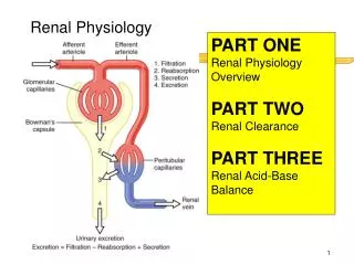
Determinants of Glomerular Filtration • The most important function of the kidney is the process of glomerular filtration. • The process of filtration is analogous to fluid movement across any capillary wall and is governed by Starling's forces. The glomerular filtration rate (GFR) is thus determined by both hydraulic and oncotic pressure differences between the glomerular capillary and Bowman's space as well as by the permeability of the glomerular membrane: • GFR = LpS - (Δhydrostatic pressure - Δoncotic pressure) • where Lp = glomerular permeability and S = glomerular surface area. • and so oncotic pressure within Bowman's space is essentially zero.
Determinants of Glomerular Filtration 1.Transglomerular (hydraulic) pressure (TGP)—the most significant determinant of GFR. the glomerular capillary is unique in that it can regulate intraglomerular capillary pressure (IGP) independent of systemic pressures through changes in afferent and efferent arteriolar tone. the pressure within Bowman's space is essentially zero 2.Renal plasma flow—is 20% but an increase in RPF leads to an increase in absolute GFR. 3.Glomerular permeability—an increase in permeability does not lead to an increase in GFR. It may, lead to increased filtration of larger molecules not normally filtered, such as albumin. Reductions in permeability or in glomerular surface area can lead to reductions in GFR. 4.Oncotic pressure—Under normal circumstances, plasma proteins are not filtered across the glomerular membrane
Regulation of Glomerular Filtration Rate • GFR is tightly maintained at a relatively constant level despite large fluctuations in systemic arterial pressures and RBF through : • 1.Autoregulation • as renal perfusion pressure increases, afferent arteriolar resistance increases, so glomerular pressure and GFR stable • if BP falls, afferent resistance falls, efferent resistance increases, and GFR and RBF preserved • mechanisms responsible for renal autoregulation not understood
Regulation of Glomerular Filtration Rate • 2.Tubuloglomerular feedback (TGF) • tubular ultrafiltrate flow rates are monitored by cells in the macula densa. • If SN-GFR increases delivery of Na+ and Cl- to the distal tubule also increases This increased Cl- delivery triggers the macula densa increase in afferent arteriolar tone and decrease RPF. • TGF is a mechanism to minimize salt and water losses through regulation of GFR. • The mediators of this response are not well understood. • Under abnormal conditions, neurohumoral responses become more important. • With significant reductions in effective circulating volume, both norepinephrine and angiotensin II maintain GFR through arteriolar vasoconstriction reduced RPF. • renal prostaglandins (PGs) and NO offset afferent arteriolar vasoconstriction
Renal clearance • The best estimate of GFR can be obtained by measuring the rate of clearance of a given substance from the plasma whitch It must: • Be able to achieve a stable plasma concentration, • Be freely filtered across the glomerulus, • Not be secreted, reabsorbed, synthesized, or otherwise metabolized by the renal tubules • Not be affected by any other means of removal from the plasma. • GFR = U[X] × urine volume/P[X]
Renal clearance • This is called the clearance of a substance and reflects the amount of plasma that is completely cleared of the substance per unit time. • The substances have been used clinically to estimate GFR: 1.Inulin. Inulin is a fructose polysaccharide, inulin clearance is the best measure of GFR. it is not clinically useful as it is difficult to administer and difficult to measure. 2.Radiolabeled compounds. These include iothalamate and diethylenetriaminepentaacetic acid (DTPA). These clearances are also very accurate but are limited in clinical use by their cost and availability 3.Creatinine. • The most widely used estimate of GFR is the 24-hour creatinine clearance (CrCl). • utilizes endogenous creatinine, which is produced at a constant rate. • The rate of production varies from individual to individual, but for a single individual daily variability is less than 10%. • easy to perform , cheap, and is available. • it is less accurate than inulin clearance, as some creatinine is cleared from plasma through proximal tubular secretion; thus, a CrCl overestimates true GFR, on average, by 10% to 20%.
Plasma Markers • simpler method to estimate GFR. • the substance must fulfill the preceding criteria. • Three substances have been utilized: 1. Plasma creatinine (PCr), the most widely used • there is marked variation in production rates between individuals, depends upon muscle mass, which is influenced by age, sex, and body mass. • the relationship of PCr to GFR is relatively constant • every 50% reduction in GFR results in a doubling of PCr. • There are limitations to the use of the PCr that should be noted: • As GFR falls, tubular secretion of creatinine increases and PCr may not change noticeably until there has been a significant drop in • Creatinine production may increase in increased muscle breakdown or with increased dietary protein intake or supplementation and lead to an underestimation of true GFR. • Creatinine production may decrease with liver cirrhosis, leading to an overestimation of true GFR.
Plasma Markers 2. Plasma urea • Urea production and excretion are highly variable, influenced by dehydration, high-protein diets, and increased tissue breakdown. • it is a much less reliable marker of GFR than is the PCr and should not be used as the sole determinant. 3. Plasma cystatin C • an endogenous protein found in all nucleated cells. • It has a constant rate of production unaffected by diet, and clearance is not influenced by tubular functions. • This test is not widely available at present but is likely to replace PCr as the standard test in GFR assessment.
Mathematical Correction. • A number of mathematical formulas have been developed to improve the accuracy of the PCr estimation of GFR. • The two most widely used are : 1.Cockcroft-Gault, originally developed from data collected from individuals with normal renal function, is a simple formula to estimate CrCl (not GFR) that corrects for age, sex, and body mass. The formula is 2. modification of diet in renal disease (MDRD) formulas. more accurate than the Cockcroft-Gault formula.:
Hormonal Control of Renal Vascular Tone • Vasoconstriction • Angiotensin II • Norepinephrine • Vasopressin • Endothelin • Atrialnatriuretic peptide • Vasodilation • Nitric oxide • Carbon monoxide • Prostaglandin E2 • Acetylcholine • Serotonin/bradykinin • Glucocorticoids
Hormonal Control of Renal Vascular Tone • Angiotensin II. • stimulates vasoconstriction of the efferent arteriole (and to afferent to lesser degree) • maintains GFR in physiologic conditions and disease states • decreases K filtration through actions on glomerularmesangium • Norepinephrine. • vasoconstricts interlobular, afferent, and efferent vessels • mesangial cell contraction • stimulates production of vasodilator PGs (ex: PGE2) • stimulates renin secretion by JGA (promoting ANGII formation)
Hormonal Control of Renal Vascular Tone • Endothelin. • Endothelin is the most potent vasoconstrictor yet identified. • three isoforms: ET1, ET2, ET3 • endothelin receptors are subclassified into: • ET(A), which are purely vasoconstrictive • ET(B), may cause either vasodilation, by stimulating the release of nitric oxide from endothelial cells, or vasoconstriction of vascular smooth muscle. • ET-1 release is stimulated by angiotensin II, antidiuretic hormone, thrombin, cytokines, reactive oxygen species, and shearing forces acting on the vascular endothelium. • ET-1 release is inhibited by nitric oxide as well as by prostacyclin and ANP • ET-1 stimulates aldosterone secretion, produces positive inotropy and chronotropy in the heart, decreases RBF and GFR, and releases ANP.
Hormonal Control of Renal Vascular Tone • AtrialNatriuretic Peptide. • ANP is a vasoactive hormone synthesized primarily by the atria in response to stretching, which occurs during physiologic levels of volume expansion • The primary actions of ANP on thekidney are increased GFR and natriuresis. • ANP can increase GFR without a change in RBF by the combination of afferent arteriolar vasodilatation and efferent arteriolar vasoconstriction. • ANP dilates vessels that have been preconstricted by norepinephrine, angiotensin II, or vasopressin. • ANP production increases during bilateral obstructive uropathy, which may be one mechanism of preserving GFR • decrease intravascular volume • increased in CHF, but renal responses attenuated in severe CHF
Hormonal Control of Renal Vascular Tone • Nitric Oxide. • Nitric oxide (NO) is a highly reactive gas that participates in multiple physiologic and pathophysiologic reactions in the body. • NO is synthesized from the reaction between arginine + NADPH + oxygen citrulline + NADP + water + NO. catalyzed by (NOS). • NOS enzymes are differ in distribution, expression, and stimuli. • Neuronal NOS (nNOS, NOS-1) and endothelial NOS (eNOS, NOS-3) are expressed and iNOS (NOS-2) is inducible. • eNOS is found in the vascular endothelium, and the NO produced there plays a key role in vasodilation and vascular remodeling. eNOS expression is stimulated by shear stress through activation of the tyrosine kinase c-Src , by heat shock protein 90 , by oxidant stress , and by vascular mediators such as bradykinin, serotonin, adenosine, ADP/ATP, histamine, and thrombin. • NO diffuses to vascular smooth muscle cellsactivates soluble guanylylcyclase (sGC)producing guanosine 3′,5′-cyclic monophosphate (cGMP)activates both cGMP- and adenosine 3′,5′-cyclic monophosphate (cAMP)-dependent protein kinases (PKG and PKA, respectively)smooth muscle relaxation. • eNOS blockade increases renal vascular resistance and decreases the glomerularultrafiltration coefficient
Hormonal Control of Renal Vascular Tone • Carbon Monoxide. • Carbon monoxide (CO) gas is a reactive diffusible mediator with multiple effects on the body and especially in the kidney. • Hemeoxygenase (HO), an essential enzyme in heme, resulting in the formation of iron, carbon monoxide, and biliverdin • Biliverdin is subsequently converted to bilirubin by biliverdinreductase. • HO is expressed in two forms, constitutive HO-2 and inducible HO-1. • Increased CO production produces vasodilation in the kidney and can counteract catecholamine-induced vasoconstriction • In particular, both HO-1 and HO-2 are highly expressed in the medulla and help maintain renal medullary blood flow • In cirrhosis, decreased renal expression of HO-1 is linked to renal dysfunction • CO also regulates sodium transport in the loop of Henle, with HO-2 blockade inhibiting sodium excretion and stimulation increasing natriuresis and diuresis • The other primary effect of CO in the kidney is renoprotection from oxidant injury. • CO has documented anti-inflammatory, antioxidant, and cytoprotective actions • a patient with a genetic HO-1 deficiency had significant tubular and vascular endothelial injury • Increased CO is protective against ischemia-reperfusion injury in native and transplant kidneys • Induction of HO-1 (bioflavonoids) protects against tubular damage and improves renal transplant function
Sodium handling by the kidney • 99% of glomerular filtrate is reabsorbed by the kidneys • PCT: • 65 – 70% of filtrated Na and water are reabsorbed by the PCT by: • Na-K ATPase in basolateral membranes Na out of the cell & K into the cell decrease intracelualar [Na] favoring entry of Na in to it from tubular lumen by Na-H antiprter on apical membrane • Cltrasported across the apical mem. in exchange for a base & exits the cell across basolateralmem. in exchange for a base • Oncotic pressure in peritubular capillaries > hydrostatic pressure favoring transport of fluid into capillaries from Intracellular spaces • So filtrates is iso-osmotic as it leaves PCT • Osmotic diuretics act in this segment
Sodium handling by the kidney • Loop of Henle • 25% of Na is reabsorbed here • Na reabsorbed in the TALH by Na-K-2Cl transporter • Loop of Henle is less permeable to water at the end the filtrate is dilute (50-60 mOsm/L) compared to plasma (300 mOsm/L) • Loop diuretics interfere with Na & Clreabsorption here • DCT • 5% of Na is reabsorbed here • Thiazide diuretics affects Na reabsorption here
Sodium handling by the kidney • Collecting tubules • 2-5 % Na & water reabsorption • Contain principal epithelial cells and the type A and type B intercalated cells • principal epithelial cells are sensetive to the aldesterone • Aldesterone increase reabsorption of Na by epithelial Na channel (ENaC) into the cell augment the amount and activity of Na-K ATPase in the basolateralmem. low [Na] in the cell favors entry of Na via ENaC from tubular lumen • Aldosterone increases K secretion by the principal cells via a distinct K channel in the apical mem. on the cell • Aldosterone facilitates increased secretion of H by increasing the activity of H-ATPase in the apical mem of the type A intercalated cells • Some intercalated cells are involved in K reabsorption • Spironolactone increase Na excretion & inhibit K secretion • Amiloride inhibit ENaC & K secretion
Water handling by the kidney • Kidneys are the major regulators of body water content: • Excessive water can be efficiently excreted by urinary dilution • In water deprivation water excretion can be minimized by urinary concentration • Urinary dilution requires that water reabsorption be inhibited which depend on: • Adequate GFR in order to deliver the filtrate to the diluting segment • Intact function of the water-impermeable nephron segment • ADH • ADH secred by the supraotpic and paraventricular nuclei in the hypothalamus posterior pituitary circulation • ADH stimulated by inceasedtonicty,hypovolemia,pain&nausia • Inhibited by law plasma osmolarity • ADH inceasespermiability of distal segment of nephron (CT) to water (20%) • ADH + V2 augment intracellular cAMP activate protein kinase A assembly of water channels (aquaporin-2) on apical membrane • In the absence of ADH impermeable to water dilute urine (minimal urineosmolality is 50-60)
Water handling by the kidney • Urinary concentration • 65-70% of filtered water is reabsorbed with Na in PCT in same proportion as they exist in the plasma fluid leaves the PCT has the same concentration of Na as plasma in DLH water leaves but not NaCl [NaCl] & tonicity is extremely high in ALH water is not permeable but salt- and urea- permeable, the absorbed NaCl enters the medullaryinterstitium & increases its tonicity, as the water remains in the lumen, the filtrate becomes dilute in collecting tubules permeability of water is under influence of ADH increase concentration • Urea diffuses & transported across the more distal collecting ducts increasing medullary tonicity • Countercurrent exchange • Water in the blood flowing in descending vasa recta moves into interstitium with higher tonicity environment in the medulla • As the blood approaches the bend of vasa recta, the [urea] & [NaCl] increased water moves back • This prevents the dilution of medullary interstitial tonicity
HCO3 handling by the kidney • Metabolic processes 1 meq/kg/day of acid that must be buffered consumming bicarbonate • So the kidney must conserve all bicarbonate filtered at glomerulous • 70-80 % reabsobed in PCT • Na/H antiport H secretion H + HCO3 carbonic acid CO2 + H2O (by carbonic anhydrase) enter lumen carbonic acid H + HCO3 enter capillary by basolateral Na- HCO3 cotranspoter
HCO3 handling by the kidney • In Collecting duct • The remainder is fully reabsorbed in collecting duct • For evey H one HCO3 is returned to plasma • Addistionaly more H secretion occurs in this segment regenerating lost HCO3 amount HCO3 returned to extracellular fluid by the kidney exceeds the amount of HCO3 filtered • The additional amount of HCO3 generated, repairs the base dificit created by the use of HCO3 in buffering the acid generated by metabolic process of the body • The H secretion in the distal nephron is an active process • HCO3-Cl exchanger in basolateral membrane HCO3 into capillary which is depends on the activity of H-ATPase in the apical mem of the typeA intercalated cells • Aldosterone increases H secretion via apical H-ATPase • The pH of the filtrate in distal lumen can be 4.5 – 5 • H secreted into lumen binds to HPO4 or NH3(ammonia, diffusable) H2PO4 (titrabe acid) & NH4(ammonium, non-diffusable)
Calcium handling by the kidney • plasma ca: -ionized -complexed with ions -protein bound • Ionic Ca and Ca complexed with other ions freely filttered by glomerulous • 70% of filltered Ca is reabsorbed in PCT • 20% absorbed in TALH • Reabsorbtion is passive depending on Na reabsorption via the paracellular pathways • 5-10% reabsorbed in DCT by an active process stimulated by PTH & Vit D • Factors that affect Ca reabsorption: volume status, PTH • Hypercalciuria predisposes to Ca stone disease
Potassium handling by the kidney • K is freely filtered and 90% is reabsorbed by PCT and ALH • Reabsorption of K is not significantly regulatable • The major mechanism by which the kidneys get rid of k excess is by means of k secretion in the collecting duct(principal cells) • K secretion is controled by: • Aldesteron • rate of fluid flow in the distal nephron • Na delivery to the distal nephron • the serum potassium concentration • the systemic acid-base status & presence of non-reabsrbable anions in the lumen of distal nephron
Uric acid handling by the kidney • Uric acid undergoes glomerular filtration, proximal tubular reabsorption by means of an urate-anion exchanger and a minor degree of proximal secretion by a proximal tubular apical voltage sensitive pathway • Uricosuric states may be associated with formation of uric acid and calcium stones • Acidic pH of tubular filtrate promotes crystallization of uric acid • Excess formation of uric acid crystal in tubules that may accompany acute tumor lysis could give rise to acute renal faliure • Hydration and alkalization of urine to pH greater than 6.5 inhibit uric acid crystallization
Endocrine functions of the kidney • Erythropoietin • secreted by tubulointerstitial cells of the kidney • Promotes amplification and maturation of erethroid precursor in the bone marrow • Vitamin D • Vit. D3 25-hydroxycholecalciferol 1,25-hydroxycholecalciferolincrease absorption of Ca + Ph in the gut
Endocrine functions of the kidney • renal angiotensinaldosterone system • Renin is secreted by the myoepithelial cells(juxtraglomerular cells) in the walls of afferent arterioles • Factors that stimulate renine secretion include • Reduced perfusion pressure in afferent arteriole • B-adrenergic stimulation via the sympathetic nerves • Factors that inhibit renin release • Increase perfusion pressure • Angiotensin II • ANP
Thank You Dr. Mohammad K. Khan