Download
1 / 33
420 likes | 813 Views
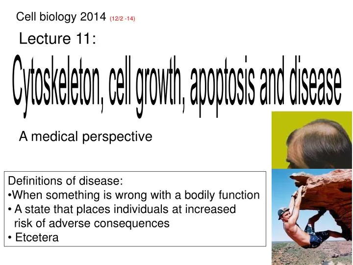
Cell biology 2014 (12/2 -14). Lecture 11:. Cytoskeleton, cell growth, apoptosis and disease . A medical perspective. Definitions of disease: When something is wrong with a bodily function A state that places individuals at increased risk of adverse consequences Etcetera.

E N D
Cell biology 2014 (12/2 -14) Lecture 11: Cytoskeleton, cell growth, apoptosis and disease A medical perspective • Definitions of disease: • When something is wrong with a bodily function • A state that places individuals at increased • risk of adverse consequences • Etcetera
The cause of different diseases Mendelian/Genetic diseases Heart disease Allergy 100% genetics 100% environment Infectious diseases Diabetes Cancer Complex multi-factorial diseases
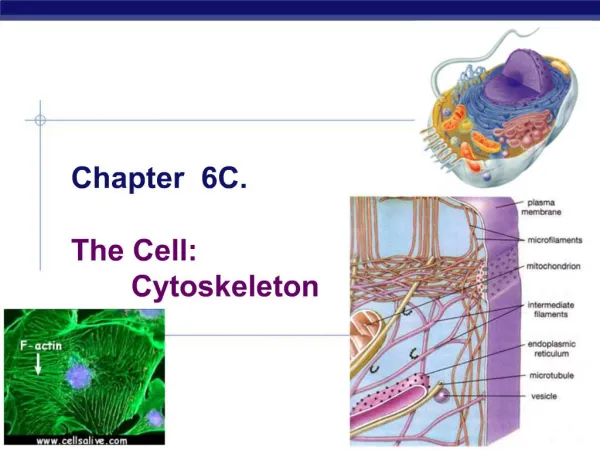
Three cytoskeleton systems: distinct but overlapping functions Intermediate filaments Actin filaments Microtubules - Cell shape and integrity - Motility of the whole cell or cellular appendages - Intracellular organization
Intermediate filaments and epithelia blistering 2. 1. Cytosolic intermediate filaments support: 1) Cell-cellcontacts (desmosomes: cadherins) 2) Cell-ECMcontacts (hemidesmosomes: integrins) 1 + 10 5 + 14 Mutations in keratin 5 or 14 cause Epidermolysisbullosa simplex (1/40 000), a disease manifested as blistering of the epidermis. Albert et al Fig. 16-21
Consequences of dysfunctional nuclear lamina Lamin A, B & C: intermediate filament proteins stabilize the nuclear envelope Laminopathies are genetic diseases manifested as either: I.Dystrophy of skeletal and/or heart muscles, caused by mutations affecting Lamin A/B or proteins attaching lamins to the nuclear envelope II.Progeria, caused by mutations in the lamin A gene, or in a lamin A processing enzyme. This result in excessive farnesylation.A farnesyltransferase inhibitor, initially developed to target oncogenic Ras, delays progression.
The actin cytoskeleton – an overview • Support of the plasma membrane • Cell migration • Contraction Muscle contraction Cytokinesis
Arp 2/3 WASP Fanta Fanta Dysfunctional actin regulation in Wiskott-Aldrich syndrome • Wiskott-Aldich syndrome (1/150.000) is caused by mutations • in the Wiskott-Aldrich syndrome protein (WASP) ZZZZ Arp 2/3 WASP • Manifested by: • Thrombocytopenia • Eczema (skin blushing) • Immunodeficiency syndrome Underdeveloped cortical actin results in defective platelets Fanta Fanta Deficient migratory and phagocytotic capacity of immune cells - Can only be cured by a hematopoietic stem cell transplant
Pathogenic E. coli: actin dependent colonization Enteropathogenic E. coli induce actin containing pedestals in intestinal epithelia Virulence factors that activate N-WASP Activation of Arp 2/3 Loss of absorptive surface is one cause for the associated diarrhea
ActA 1. 2. 3. 4. 5. ActA ActA Listeria: actin dependent motility 1-3) Phagocytosis and escape from phagosome 4) Bacterial multiplication 5) Penetration of a neighboring cell through actin based motility Arp 2/3 video 24.3-listeria_parasites
The microtubule system - an overview ER • Intracellular • organization Golgi Organelle positioning Chromosome segregation Chemotactic agent - Cell motility Cell polarisation and transport Movement of cellular appendages
Lissencephaly: defective neuron migration Lis1 • Lissencephaly ("smooth brain," 1/30.000) is a disorder characterized by the lack of normal convolutions (folds) in the brain Lissencephalybrain Normal brain mutated in many cases loss of dynein ( ) dependent centrosomereorientation defective cell polarization - 1/30 000 births, early death in severe cases
Non-functional cilia in Kartagener syndrome • Kartagener syndrome (1/20.000) is caused by mutations • affecting cilia specific dynein • - Manifested by respiratory infections, infertility and situsinversus Upper respiratory epithelia Patient with Kartagener syndrome Normal Goblet cell Bacteria is caught in mucus and cleared by a cilia mediated flow Bacteria is not cleared due to defective cilia
Microtubule-poisoning drug: Taxol • Alkaloid ester isolated from the bark of • Taxus brevifolia (Pacific yew) • Stabilization of microtubules Therapeutic uses: Treatment of breast, lung and ovarian cancer Prevention of restenosis of coronary stents (Surface coating of stents local action) Major side-effects: Bone marrow suppression, gastro-intestinal upset and peripheral neuropathy
Microtubule-poisoning drug: Vinca alkaloids • Isolated from Catharanthusroseus • Named: Vinblastine, Vincristine,Vindesine • and Vinorelbine • Sequesters tubulin Therapeutic use: Treatment of leukemia, lymphoma, breast, lung, prostate, skin and testicular cancer Major side-effects: Bone marrowsuppression, gastro-intestinal upset and peripheralneuropathy
An oncology perspective on signal transduction, cell growth, checkpoints, apoptosis and the cytoskeletons X X X X X X X X X X X X Progression towards malignancy involves : i) uncontrolled proliferation, ii) resistance to apoptosis, iii) cell migration, iv) tissue invasion • Clonal evolution • Selection of • malignant clones Metastasis animation 20.2 -contact_inhibition video 20.1 -breast_cancer_cells
Two distinct types of ”cancer genes” Oncogenes Tumor suppressors On On Gene X Gene Y Off Gene X Gene Y Off On On Off X On Gain-of-function Loss-of-function A single genetic change Two independent events Dominant phenotype
CKI Ras Bcl-2 Rb Definitions: oncogenes and tumor suppressors An oncogene is a gene that when mutated, or overexpressed, contributes to converting a normal cell into a tumor cell (constitutive activity dominant phenotype) Onc point mutation overexpression A tumor suppressor-gene is a gene whose loss, or inactivation, contributes to converting a normal cell into a tumor cell (recessive phenotype) TS p53 Inactivating point mutations or loss of the entire gene (germ line mutation in one allele and/or acquired somatic mutations)
Cell type specific proliferative signals Cells from different tissues express distinct sets of growth factor receptors and signaling proteins Cell type B Cell type C Cell type A Major mitogen signaling pathway: RTK Wnt Hedgehog Alterations in tumors:RTK signals Wntsignals Hedgehog signals
Wnt RTK Frizzled Ras APC Aberrant proliferative signals in tumors XGF Hedgehog Onc Onc TS Patched Smoothened Onc Dishevelled TS TS Raf Axin GSK-3b Fused Onc SuFu Erk TS b-catenin Onc myc Onc myc G1 Gli Gli Onc Onc Onc Onc myc Onc Onc G1 G1
P P Cdk Cdk G1 S TGF-b DNA replisome Insensitivity of tumors to anti-growth signals Mitogen signaling p15 p21 p16 TS TS viral The retinoblastoma pathway HPV E7 Onc Rb E2F Cdc6 ORC = germ line mutations identified
p53 BH3 only TS TS Onc Bax Bcl-2 Cyt. C Evading cell death (apoptosis) Ligand TS Survival factor signaling Onc Death receptor TS Adaptor Caspase 8 Caspase 9 Caspase 3 Apoptosis
Randomly acquired oncogenic mutations drives tumor progression Mutation 1. Self-sufficiency in proliferative signals 1 2. Insensitivity to anti-growth signals 2 3. Evading cell death (apoptosis) 3 4. Limitless replicative potential 4 5. Sustained angiogenesis 5 6. Metastasis capability 6
Same “diagnosis” but different set of mutations Patient A with diagnosis X 1 2 3 4 5 6 X X X X Patient B with diagnosis X 1 2 3 4 5 6 X X X
How many somatic mutations during a life time? Cell death and replacement risk for mutations & chromosomal instability Year 1-15 Controlled and co- ordinated divisions Uncontrolled divisions Tumors 1013 – Human diploid genome: ~6 x10⁹bp – Only some few errors per replication cycle – Average t½ of cells is 7 years (range: 24h to >100 years)
Normal cells have a very low rate of mutations Cancer related genes Other genes Random mutation Time Due to the low normal mutation frequency, progression to a fully malignant tumor is statistically improbable How come that malignant tumors have either a lot of mutations (~10 %) or chromosomal aberrations (90 %)? (~400 genes are frequently altered in tumors, 6 to 80 genes per “patient”)
Genomic instability: Two distinct levels 1. Defective DNA repair (MIN) Mutation in a gene encoding some enzyme required for DNA-repair X X No repair many mutations accelerated tumor progression 2. Chromosome segregation errors (CIN) Mis-segregation due to a defective a gene that encodes some protein essential for high fidelity chromosome segregation
1 2 3 4 5 6 MIN reflects an escalated mutation rate Cancer related genes Other genes DNA repair(TS) Genetic alteration Time ?? Often uncertain which ones of all the mutations that contribute to tumor progression MIN: mini-satellite DNA instability (due to defective DNA repair)
CIN through excessive centrosomes Two centromes More than two centrosomes Kinetochore attachments satisfy the spindle checkpoint miss-segregation aneuploidy CIN: Chromosomal instability
APC CIN through loss of APC The tumor suppressor gene product APC functions as a MT plus-end stabilizing protein ( ) that facilitates stable MT-kinetochore connections Satisfied spindle checkpoint Centromere Centrosome AC Kinetochore CIN: Chromosomal instability
CIN through a defective spindle checkpoint 1) A normal cell Delayed anaphase until all kinetochores are attached 2) A tumor cell with a (partially) defective spindle checkpoint
Cancer x x Genomic instability and tumor progression A stable genome Too much genetic instability ”Optimal” genetic instability ”Selection barriers”
Principles of cancer treatment Surgery- Impossible to remove all cancer cells Radiation- Targets both cancer- and normal cells Chemotherapy- Side-effects • General chemotherapy: drugs that interferes with: • DNA-replication • DNA structure • The function of the microtubule-system • Chemotherapy may also include cell type specific drugs. E.g. inhibition of hormone dependent tumor growth
x x Selective killing of tumor cells by chemotherapy Mutations that inactivate various checkpoints are common in malignant tumors no cell cycle arrest in patients treated by chemotherapy!