Download
1 / 45
1.19k likes | 3.69k Views
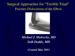
Surgical Approaches for “ Terrible Triad ” Fracture-Dislocations of the Elbow. Michael J. Medvecky, MD Seth Dodds, MD Created May 2011. What is a Terrible Triad?. Elbow dislocation Coronoid fracture Radial head fracture. Terrible Triad Injuries: Mechanism of Injury.

E N D
Surgical Approaches for “Terrible Triad”Fracture-Dislocations of the Elbow Michael J. Medvecky, MD Seth Dodds, MD Created May 2011
What is a Terrible Triad? • Elbow dislocation • Coronoid fracture • Radial head fracture
Terrible Triad Injuries: Mechanism of Injury • Fall on an outstretched hand • Axial load • Relative elbow extension • Valgus • Forearm rotation • Supination The ultimate “Posterolateral rotatory instability”
Terrible Triad Fracture-Dislocation • What is so terrible about it? • Extremely unstable • Loss of joint congruency • Instability • Fracture fragments are usually quite small • Difficult to repair • Patients don’t routinely do “well” • Unaware of the magnitude of the injury for the elbow • Residual instability • Stiffness
Lateral Collateral Ligament • Radial collateral ligament • Lateral ulnar collateral ligament • Annular ligament
Medial Collateral Ligament • Anterior bundle • Posterior bundle • Transverse bundle
Anterior capsule Brachialis Anterior bundle of MCL Anteromedial facet of coronoid Fx propagation into this region may cause functional MCL incompetancy Proximal Ulna - Anterior Coronoid
Posterior dislocation & radial head fracture Injury Patterns
Posterior dislocation & radial head fracture Posterior dislocation, radial head & coronoid fractures “Terrible Triad” Injury Patterns
Posterior dislocation & radial head fracture Posterior dislocation, radial head & coronoid fractures “Terrible Triad” Transolecranon fracture-dislocations Anterior Posterior Injury Patterns
Terrible Triad InjuriesPatient and injury assessment • Patient evaluation • Associated injuries • Mechanism of injury • Soft tissue status • Radiographs (possible traction views) • Post-reduction CT w/ 3D recons • Operative timing • As urgently as possible but during the daytime • Pre-op planning for appropriate equipment
Type I: nondisplaced No block to forearm rotation, displacement < 2mm Type II: displaced Internal fixation possible Type III: displaced, severely comminuted Judged to be irreparable Type IV: fracture + dislocation Radial Head Fractures:Modified - Mason Classification
Regan & Morrey Type 1 tip Type 2 < 50% May be stable Type 3 > 50% usu very UNstable Classification: Coronoid Fractures
O’Driscoll Classification Type I: tip Type II: anteromedial facet Type III: base Classification: Coronoid fractures
36 consecutive patients treated: Fix or suture coronoid Repair / replace radial head Repair LCL If still unstable, repair MCL If still unstable, hinged ex-fix Terrible Triad –Treatment ProtocolMcKee, Pugh, Schemitsch,et al JBJS(A) ‘04
What’s injured? Radial head only Radial head type 1 coronoid Radial head type 2 or 3 coronoid Proximal ulna / olecranon Medial Approach Needed if: plate coronoid fracture transpose ulnar nerve repair or reconstruct MCL Surgical Planning: Approaches Radial head replacement & common proximal ulna fracture exposes coronoid tip
3 steps: Repair radial head Secure radial head to the radial neck Avoid impingement of plates during forearm rotation. Small K wires used provisionally. “mini-fragment” screws (1.5 to 2.7 mm), countersink heads Secure radial head to neck with 2.0 or 2.7 L-shaped plates or mini blade plates Internal fixation
Comminuted Radial Head FractureRole of the Radial Head Arthroplasty • Excision will lead to instability • Functional spacer • Creates stability by increasing radial length & restoring valgus restraint
Medial Epicondyle FCU Ulnar Nerve Terrible Triad: Medial Instability ? • Repair MCL • Reconstruct through bone tunnels • Suture Anchors • Palmaris autograft or allograft tendon • Repair muscle origins Pronator FCU Medial Epicondyle Nerve Ulnohumeral joint reduced
Terrible Triad: Persistent Instability ? Uniplanar Lateral Frame • Hinges Multiplanar Compass Hinge
Positioning: supine vs lateral Supine: Better access and visualization of anterior joint & coronoid Lateral facilitates ulnar length, lessens needs for assistants Surgical approach: Midline Posterior Kocher (posterolateral) vs Kaplan (anterolateral) Anteromedial Posteromedial Percutaneous coronoid fixation Surgical Planning
Anconeus – ECU interval Lateral: Kocher Approach
Anterior column exposure Supracondylar ridge Anterior to mid-axis of radiocapitellar joint Utilize LCL tear Incise anterior capsule Exposes anterior coronoid Replacement or fixation Lateral: Kaplan Approach
Lateral Approach: Deep dissection • Access to anterior ulno-humeral joint • Elevate the extensors • Stay superior to the LCL • Able to visualize the PIN • Arthrotomy • Release of the lateral capsule and annular ligament
Medial supracondylar ridge Pronator teres - brachialis interval Incise anterior 1/2 flexor-pronator mass Anterior capsule Anteromedial Approach to Coronoid
Medial supracondylar ridge Pronator teres - brachialis interval Incise anterior 1/2 flexor-pronator mass Anterior capsule Anteromedial Approach to Coronoid
Medial supracondylar ridge Pronator teres - brachialis interval Incise anterior 1/2 flexor-pronator mass Anterior capsule Anteromedial Approach to Coronoid
Exposure of: Coronoid Sublime tubercle MCL Proximal ulna MCL reconstruction or repair ORIF AM facet of coronoid Buttress plating of coronoid Posteromedial Approach to Coronoid
Necessitates ulnar nerve exposure and transposition Palpate sublime tubercle Incise FCU ulnar attachment distal to sublime tubercle and proceed proximally -> anterior bundle of MCL. Posteromedial Approach to Coronoid
Terrible Triad Injuries: Rehab • Rehab • Stiffness vs. Instability • Cautious • Posterior splint • 14 days post-op • Cuff and collar • Guided rehab is essential • Flexion first! • Active and passive • Active and passive forearm rotation at 90° • Begin extension at 3 weeks, active only • Start supine—active against gravity
Terrible Triad Injuries: Summary • Not so Terrible • Isolated injury & cooperative patient • Stable repairs & motion • Coronoid fixation • Radial head arthroplasty vs. ORIF • LCL repair • Terrible • Poor stability after repairs complete • Multi-trauma • ICU stay • Head injuries • Non-weight bearing on lower extremities • Uncooperative patient
Conclusions If you would like to volunteer as an author for the Resident Slide Project or recommend updates to any of the following slides, please send an e-mail to ota@aaos.org E-mail OTA about Questions/Comments Return to Upper Extremity Index