Download
1 / 49
500 likes | 579 Views
Explore the anatomy and function of the autonomic nervous system, its divisions, motor innervation to muscles and glands, neurotransmitters involved, and differences between the parasympathetic and sympathetic divisions.

E N D
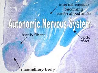
Biology 220 Anatomy & Physiology I Unit IXAUTONOMIC NERVOUS SYSTEM Chapter 14, pp.513-529 E. Gorski/ E. Lathrop-Davis/ S. Kabrhel
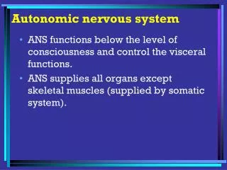
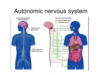
General Information • VISCERAL MOTOR INNERVATION • Motor innervation to smooth muscle, cardiac muscle and glands • Consists of two opposing divisions • most structures receive innervation by both divisions (= dual innervation) • helps ensure stability of internal environment • involuntary activity (subconscious)
Somatic Autonomic Effectors skeletal muscle cardiac, smooth muscle; glands Peripheral Pathway 1 motor neuron 2 motor neurons (preganglionic & postganglionic) Fiber Type(s) A B (preganglionic) C (postganglionic) Motor Ganglia none autonomic (chain/prevertebral or terminal) Neuro-transmitter(s) ACh (always excitatory) ACh or Norepinephrine (excitatory or inhibitory depending on receptors) Comparison of Somatic and Autonomic Nervous Systems
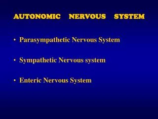
Divisions of theAutonomic Nervous System (ANS) • Parasympathetic Division (PD) -- primarily concerned with gaining and conserving energy • “rest-repose” (resting and digesting) system • e.g., enhances digestion, slows heart rate • Sympathetic Division (SD) -- primarily concerned with responses to life-threatening situations • “fight-or-flight” system • e.g., inhibits digestion, raises heart rate
General Structure In both divisions, 2 motor neurons involved: preganglionic and ganglionic • preganglionic neuron - has its cell body in gray matter of brain or spinal cord • axon extends out via cranial or spinal nerve • small diameter, lightly myelinated (type B) fibers • release ACh at synapse to excite ganglionic neuron (= postsynaptic membrane)
General Structure (con’t) • ganglionic neuron – cell body is in autonomic ganglion • axon (postganglionic axon or fiber) is type C and extends from autonomic ganglion to effector • postganglionic axon releases either • norepinephrine - released by most fibers of sympathetic division • ACh - released by fibers of parasympathetic division • action of neurotransmitter is excitatory or inhibitory depending on receptors in effector
Differences in Structure Between Divisions 1. Site of origin 2. Location of ganglia 3. Length of preganglionic and postganglionic fibers See Table 14.1, p. 516
1. Site of Origin • parasympathetic • cell bodies of preganglionic neurons are in brain stem nuclei or gray horns of sacral spinal segments • fibers of preganglionic neurons exit from brain via cranial nerves or sacral segments of spinal cord (= “craniosacral outflow”) • sympathetic • cell bodies of preganglionic neurons are in lateral gray horns of thoracic or lumbar segments • fibers of preganglionic neurons are part of thoracic and lumbar spinal nerves (= “thoracolumbar outflow”)
2. Location of Ganglia and 3. Length of Fibers 2. Location of ganglia • parasympathetic division has ganglia generally near the structures they innervate • sympathetic division -- most ganglia lie close to the vertebral column 3. Length of fibers • parasympathetic division (generally) has long preganglionic fibers and short postganglionic fibers • sympathetic division has short preganglionic fibers and long postganglionic fibers
Comparison of Divisions Fig. 14.2, p. 514
Parasympathetic (Craniosacral) Division • preganglionic neurons • bodies in brain stem nuclei (for cranial nerves) or in gray horns of sacral spinal cord segments S2-S4(for sacral nerves) • fibers are long • release ACh at synapse • ganglia are located near structures they innervate • terminal ganglia (= intramural ganglia) • postganglionic fibers • generally short • release ACh at neuroeffector junction with effector • Several cranial nerves (III, VII, IX and X) contain parasympathetic fibers
Cranial Outflow: CN III 1. Oculomotor nerve (III) • nerve innervates smooth muscle that controls iris - causes pupil to constrict • innervates smooth muscle of ciliary body - causes lens to change shape for near vision • preganglionic fibers originate in midbrain • postganglionic fibers originate in ciliary ganglia Fig. 14.4, p. 517
Cranial Outflow: CN VII 2. Facial nerves (VII) • stimulate nasal and lacrimal glands (tearing), lower salivary glands • preganglionic fibers originate in pons • postganglionic fibers originate in ganglia in face (pterygopalatine and submandibular) Fig. 14.4, p. 517
Cranial Outflow: CN IX 3. Glossopharyngeal nerves (IX) • stimulate parotid salivary glands • preganglionic fibers originate in medulla • postganglionic fibers originate in otic ganglia within cranial cavity Fig. 14.4, p. 517
Cranial Outflow: CN X 4. Vagus nerves • major nerves (90% of craniosacral preganglionic fibers) providing parasympathetic innervation to structures of thoracic and abdominal cavities • preganglionic fibers originate in medulla • postganglionic fibers arise from terminal (intramural) ganglia located on/in target organs. • fibers from right and left intermingle to form plexuses in thoracic cavity and abdominal cavity
Cranial Outflow: CN X Thoracic Plexuses • Cardiac plexus supplies innervation to heart • Pulmonary plexus supplies innervation to lungs (bronchi, bronchioles) • Esophageal plexus supplies innervation to lower esophagus (tube through which food passes on way to stomach) Fig. 14.4, p. 517
Cranial Outflow: CN X Abdominal plexus (aortic plexus) supplies innervation to most of the digestive system (stomach, gall bladder, liver, small intestine, pancreas, proximal part of large intestine) and urinary system (kidneys) Fig. 14.4, p. 517
Sacral Outflow • preganglionic fibers arise from neurons in gray horns of sacral segments S2-S4 • axons run through ventral roots of sacral spinal nerves • postganglionic fibers arise from ganglia near organ innervated • provide parasympathetic innervation to pelvic organs • distal (last part) large intestine (including rectum and anal canal) • reproductive system (including uterus, external genitalia) • urinary bladder and ureters
Sympathetic (Thoracolumbar) Division • innervates same organs as parasympathetic - BUT also other structures not innervated by parasympathetic • sweat glands and arrector pili muscles of skin (make hairs stand up) • adrenal medulla • blood vessels • preganglionic neurons have cell bodies in lateral gray horns of thoracic or upper lumbar spinal cord segments (T1-L1) • release ACh at synapse with ganglionic neuron
Sympathetic (Thoracolumbar) Division • postganglionic fibers arise from ganglionic neurons in ganglia close to spinal cord • chain (paravertebral) ganglia are located adjacent to vertebral column (paired) • prevertebral ganglia are located anterior to vertebral column (single) Fig. 14.5, p. 519
Sympathetic Preganglionic Neurons • preganglionic fibers leave through ventral roots of thoracic or lumbar spinal nerves only • myelinated fibers pass through white ramus communicans* to reach chain ganglion (paravertebral ganglion) lying laterally to vertebrae • paravertebral chain ganglia form sympathetic chain (run from cervical to sacral regions) • typically, 23 ganglia in each sympathetic chain (23 pairs) *plural is “rami communicantes”
Sympathetic Preganglionic Neurons (con’t) • once it has reached ganglion, preganglionic fibers can: 1. synapse with ganglionic neuron in chain ganglion 2. go up or down through sympathetic trunk to synapse in chain ganglion at another level Fig. 14.6, p. 520
Sympathetic Preganglionic Neurons (con’t) 3. pass through the chain ganglion and go out to prevertebral ganglion (anterior to vertebral column near aorta - major artery along back wall) • preganglionic fibers help form splanchnic nerves • synapse with ganglionic neurons in prevertebral ganglia Fig. 14.6, p. 520
Sympathetic Innervation • paravertebral postganglionic fibers innervate: • head - eye (dilate pupil); inhibit salivary glands, lacrimal gland, nasal glands • lungs - dilate airways and blood vessels • heart - increase rate and strength of contraction • prevertebral postganglionic fibers innervate: • GI organs - inhibitory (decreases activity in liver, stomach, intestines, etc.) • urinary system - decreases output and relaxes bladder • reproductive organs • MOST postganglionic neurons release norepi *See Table 14.4
Adrenal Medulla • part of endocrine system • releases hormones: • norepinephrine - 20%, vasoconstriction • epinephrine - 80%, control of heart and metabolism • arises from same embryonic tissue as peripheral nervous system • same as postganglionic fibers of SD/ANS: acts like one big postganglionic fiber only releases “NT” into blood instead of to specific effectors • stimulated by preganglionic fiber of SD
Physiology of Autonomic Nervous System (ANS) Fibers and Receptors For: A. Parasympathetic Division B. Sympathetic Division
Fibers and Receptors: PD/ANS • postganglionic fibers are cholinergic - release acetylcholine (ACh) at effector • effectors have cholinergic receptors (bind ACh) 1. Nicotinicreceptors • direct acting • always excitatory • found dendrites/cell bodies of all ganglionic neurons (both divisions) • also found in • skeletal muscle (part of somatic nervous system) • adrenal medulla (innervated by preganglionic fibers of SD) See Table 14.3, p. 524
Fibers and Receptors: PD/ANS 2.Muscarinic receptors • indirect acting • excitatory or inhibitory • found in: • all effectors innervated by postganglionic neurons of PD • e.g., cardiac muscle - inhibited (slows heart rate) • e.g., smooth muscle and glands of GI tract - stimulated* (increases activity) • some effectors innervated by SD • e.g., eccrine sweat glands - stimulated • e.g., blood vessels in skeletal muscle - inhibited (vasodilation) See Table 14.3, p. 524
Fibers and Receptors: SD/ANS • most postganglionic fibers are adrenergic (release norepinephrine = NE) 1. Alpha receptors (): • 1 - almost all sympathetic target organs except heart • constricts peripheral blood vessels and GI sphincters • dilates pupils of eyes by acting on iris See Table 14.3, p. 524
Fibers and Receptors: SD/ANS • 2 - inhibits NE release (binds to adrenergic axon terminals); stimulates blood clotting • some sympathetic postganglionic fibers that innervate sweat glands (in skin), some blood vessels (skeletal muscles, brain, external genitalia) secrete ACh and are, therefore, cholinergic See Table 14.3, p. 524
Fibers and Receptors: SD/ANS 2. Beta receptors (β): β1- generally stimulate: • heart (increases rate and strength of contraction) • kidneys (stimulates renin secretion [long pathway resulting in water retention and increased blood pressure - will study in AP II]) See Table 14.3, p. 524
Fibers and Receptors: SD/ANS 2. Beta receptors (β): • β2 – mainly inhibition (smooth muscle relaxation) • lungs (smooth muscle of bronchi & bronchioles) • heart blood vessels (vasodilation) • many other sympathetic organs • stimulates secretion of insulin • β3– brown adipose tissue - stimulates hydrolysis (lipolysis) of stored fat - thermogenesis
Effects of Selected Drugs • Atropine - anticholinergic (blocks effects of ACh by blocking muscarinic receptors [blocks parasympathetic]) • suppresses salivation and respiratory secretions • used to dilate pupils for eye exam • dilates bronchi when inhaled
Effects of Selected Drugs • Neostigmine - anticholinesterase • prevents breakdown of ACh at neuromuscular junction with skeletal muscle, thus allowing ACh to accumulate to sufficient levels at synapse • used in treatment of myasthenia gravis (autoimmune disorder in which ACh receptors of skeletal muscle are damaged) • does not cross blood-brain barrier
Effects of Selected Drugs • Tricyclic antidepressants • prolong activity of NE and serotonin (prevent reuptake) on postsynaptic membranes within CNS --> feel good • also anticholinergic effects (cause sleepiness)
Effects of Selected Drugs • Ephedrine, phenylephrine • sympathomimetic (mimic sympathetic effects) • both used to treat colds, allergies • phenylephrine – used to treat shock; used to treat nasal congestion (Neo-Synephrine) • stimulate alpha-1 receptors, which inhibit nasal and lacrimal secretion through peripheral vasoconstriction • ephedrine also causes bronchodilation and increases heart rate • phenylephrine used to treat shock due to strong action on blood vessels
Effects of Selected Drugs • Beta blockers - block β1 receptors of cardiac muscle • reduce heart rate • treat arrhythmias (errors in heart rhythm) • Phentolamine - alpha blocker (1) used to treat hypertension (increased blood pressure) • used when hypertension is associated with excess sympathetic activity, as in when patient is also taking sympathomimetic drugs (e.g., Ephedrine, Phenylephrine)
Interactions of ANS Divisions • most organs receive dual innervation • exceptions include: • adrenal medulla • almost all blood vessels • eccrine sweat glands • normally, one division dominates • e.g., parasympathetic activity normally keeps heart rate lower • in “emergency”, sympathetic overrules
Sympathetic and Parasympathetic Tone • tone arises from near constant output of sympathetic or parasympathetic impulses • sympathetic tone used to maintain vasomotor tone (partial constriction) of blood vessels • parasympathetic tone • keeps heart rate lower • maintains partial contraction of GI tract organs (stomach, intestines) • maintains partial contraction of urinary system tubes (ureters)
Cooperative Effects of ANS Divisions • usually antagonistic, but work together to accomplish certain goals,e.g., • parasympathetic impulses cause vasodilation of blood vessels in genitalia (essential to erection - penis/clitoris), • sympathetic impulses essential to ejaculation
Unique Roles of SD/ANS • SD provides only innervation to: • adrenal medulla • sweat glands • arrector pili muscles • most blood vessels • thermoregulation (maintenance of body temperature) - depends on SD which directs blood flow • release of renin from kidney (important to fluid balance and blood pressure) • metabolic effects (increases metabolism)
Local Versus Diffuse Effects • Parasympathetic - short-lived, local effects • ACh acts indirectly at muscarinic receptors (smooth and cardiac muscle and glands) • ACh quickly hydrolyzed to acetyl CoA and choline by acetylcholinesterase (enzyme) • one preganglionic fiber synapses with only one or a few postganglionic fibers
Local Versus Diffuse Effects • Sympathetic - longer lasting, more diffuse • NE works indirectly through second messenger • takes longer, but also lasts longer • NE not hydrolyzed, but taken back up into axonal terminals of postganglionic fibers • preganglionic fibers synapse with many postganglionic fibers (divergence), often at several levels • activation of adrenal medulla results in release of epinephrine and norepinephrine
Control of ANS Function:Brain Stem & Spinal Cord • Medulla oblongata has most direct effects • centers for heart rate, vasomotor tone, respiratory system (dilate airways and blood vessels) • center for GI tone • Pons - respiratory center influences respiratory centers in medulla • Midbrain - reflex centers controlling pupil size • Spinal cord - reflexes for urination, defecation, erection, ejaculation • can be overridden by conscious controls
Control of ANS Function:Hypothalamus • exerts overall control • influences motor activity through reticular formation • also forms important part of limbic system controlling emotions • contains centers (groups of neurons with related functions) for: • heart activity (rate and force) • blood pressure • body temperature
Control of ANS: Cortical Controls • biofeedback - learned access of parasympathetic division • learn to lower heart rate, blood pressure through use of devices that give feedback on status of some part • concentrate on thoughts that calm, relax
Review of Upper Control Fig. 14.9, p. 528
Visceral Reflex Arcs Same basic elements as somatic reflex arc • receptors in visceral organs (e.g, heart, blood vessels) respond to • chemical changes, stretch, irritation • visceral sensory neurons carry impulse toward CNS • integration centers within CNS • visceral motor neurons- 2 neuron pathway - preganglionic and ganglionic neurons [postganglionic fiber]) • visceral effector (smooth or cardiac muscle) Fig. 14.7, p. 522
Referred Pain • sensory neurons of visceral pain travel same pathways as somatic sensory neurons serving nearby areas • referred pain occurs when visceral stimuli are perceived as somatic (more common pathway) Fig. 14.8, p. 523