Download
1 / 27
270 likes | 617 Views
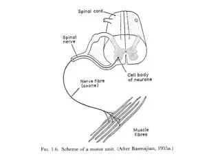
Depolarization of Muscle Membrane. The EMG signal is derived from the depolarization of the muscle membrane. The electrodes record the sum of all of the muscle fiber action potentials from all the active motor units that pass through the recording zone of the electrodes. EMG Signal.

E N D
Depolarization of Muscle Membrane The EMG signal is derived from the depolarization of the muscle membrane. The electrodes record the sum of all of the muscle fiber action potentials from all the active motor units that pass through the recording zone of the electrodes
EMG Signal • The EMG signal represents the sum of all the muscle fiber action potentials from all of the active motor units that passes through the recording zone of the electrodes.
Motor Unit Synchronization Un-Synchronized Action Potentials Reduce EMG Amplitude. Shaded areas indicate where the positive phase of one action potential overlaps in time with the negative phase of another action potential leading to the cancellation of the surface EMG (shown as the sum) [From Yao (2000) #1996] Synchronized action potentials result in an increase in the surface EMG. (shown as the sum) [From Yao (2000) #1996]
Effects of Motor Unit Synchronization Upon EMG Amplitude and Force Synchronization increases the EMG amplitude and the variability of the force. [From Yao (2000) #1996]
Effects of Synchronization Upon the Median Frequency of the EMG Signal Motor unit synchronization lowers the median frequency of the surface EMG signal. [From Yao (2000) #1996]
In general, there is a linear relation between EMG and force.
Relationship Between EMG Signal and Motor Unit Force • The EMG signal of a ST motor unit can be described as low amplitude and long duration. • The EMG signal of a FT motor unit can be described as high amplitude and short duration. • When an ST motor unit fires, it yields a relatively small amount of force with a longer time to peak force, when compared to a FT motor unit. • Since FT units are highly fatigable, the CNS is very protective of them. FT units are recruited when a rapid force is needed or only at high force levels.
Size Principle (Recruitment Threshold) • Small [Slow Twitch, Type I] are recruited first • Intermediate [Type IIa] are then recruited • Large [Fast Twitch IIb] are recruited last • The vertical lines indicate when a motor unit has fired. When the lines are closer together, the motor unit has increased its firing rate. • Fast twitch units are the first to be de-recruited, followed by intermediate and finally slow twitch units. • Notice that motor units are recruited and de-recruited at about the same force level. FT motor unit ST motor unit
Motor Unit Recruitment Motor units are recruited according to size (I, IIa, IIb). However in ballistic contractions the high threshold (MUP 2) units may show up in the EMG signal first due the faster axonal conduction velocity [see Desmedt #1994].
Electromechanical Delay (EMD) • EMD is the delay between the start of EMG and the start of force. • The electrical event, depolarization of muscle membrane (EMG) occurs before the mechanical event (force). • The delay comes from time needed to: • Propagate the action potential along the T-Tubule system • Release of calcium from the sarcoplasmic reticulum • Formation of Actin-Myosin crossbridges • Stretching of the series elastic element (SEC): titin, myosin head and arm, tendon • EMD times are effected by: level of force, muscle length, type of contraction (isometric, concentric, eccentric) , premotor state of the muscle
[From Herzog #2318] Electromechanical Delay in Gait The EMG signal shown above is from the soleus of a cat during gait. The force was measured directly from the achilles tendon. Notice that the EMG starts about 70 ms prior to force and that the EMG ends about 70 before the force ends.
Muscles have two mechanisms to modulate force: recruitment and firing rate. Depending upon the muscle and the force requirements different recruitment-rate patterns can be observed. Muscle A is the soleus, Muscle B is the bicep. By 60% of MVC the soleus has recruited all of its motor units and must rely upon increase firing rate to increase force above 60%. The bicep continues to recruit additional FT motor units until about 80% MVC.
Different firing rate control strategies: In Figure A units increase their firing sharply after being recruited, show a plateau, and then increase sharply and the higher force levels. In Figure B, unit A reaches max firing immediately after being recruited, units B and C display linear increase in firing rates.
EMG – Fatigue at 40% MVC Recruit FT Contraction begins with ST and INT FT fatigues INT fatigues FT 1 FT 2 INT 1 INT 2 ST 1 ST 2
When motor units fatigue they fire in shorter bursts and rest longer between subsequent bursts EMG – Fatigue at 80% MVC FT 1 FT 2 INT 1 INT 2 ST 1 ST 2
A single Actin-Myosin bond yields less force concentrically (1-2pN) than eccentrically (N*3-4pN), as a result, concentric EMG has a larger amplitude than eccentric EMG. The increased EMG amplitude observed in concentric contractions is due to some combination of increased recruitment and/or increased firing rate of currently active motor units. [#1845]