Download
1 / 38
390 likes | 584 Views
Intestinal Ischemia. Academic Half Day Dean Soulellis Gastroenterology. Intestinal Ischemia. Intestinal ischemia is a bunch of different pathologies involving different organ within the GI tract Best to go anatomically Celiac trunk. Celiac Trunk/Axis.

E N D
Intestinal Ischemia Academic Half Day Dean Soulellis Gastroenterology
Intestinal Ischemia • Intestinal ischemia is a bunch of different pathologies involving different organ within the GI tract • Best to go anatomically • Celiac trunk
Celiac Trunk/Axis • Supplies the lower esophagus, stomach, D1, D2, sometimes D3, and liver/pancreas
SMA • D2/D3, Jejunum, Ileum, cecum and ascending colon, and most of transverse colon
IMA • Distal transverse colon to the proximal rectum
Causes of Ischemia • Lack of blood flow • Lack of Oxygen • Thrombus (A or V) • Embolus (A or V) • Supply-demand mismatch (low flow states)
Intestines Protect Themselves • Can tolerate 75% reduction in blood flow for up to 12 hours • At any moment in time, 1 in 5 capillaries are open • Able to extract oxygen efficiently in times of need
Irreversible Ischemia • Eventually vasodilation of residual capillaries overwhelmed by ischemia • Leads to vasoconstriction and necrosis • Reperfusion injury
Intestinal Ischemia • Mechanism is familiar to you • Same thing occurs in the heart (thrombus or low-flow state), kidneys (thrombus or embolism or low-flow state), brain (thrombus or embolism), extremities, etc.
Clinical Features - Acute • Severe acute abdominal pain • Patient feels like vomiting • The problem is usually arterial – embolus, thrombus, or low-flow state
Older Patients • Often more indolent presentation – chronic thrombus formation in one of the main branches • Possible cardiac embolic event • Maybe painless in very elderly
Older Patients • A third of the very elderly will present with confusion alone! • IF painless with blood per rectum, might be low-flow state to the colon – NOMI (not “ischemic colitis”)
Younger Patients • Usually arterial embolic • More violent presentation • Think vasoactive street drugs and arrhythias
Chronic Pain • Consider mesenteric VENOUS thrombosis • Conceptually similar to DVT • Results in ongoing abdominal pain, more chronic • Ask about history of DVT, hypercoagulable states, vasculitis, previous abdominal surgery or infection
Physical Exam • Assess vitals • Watch for unusual presentation in the elderly • Abdomen may be benign early on, then progress to tender, then rigid • Distention is a very bad sign
Physical Exam • Look for bloody stool on rectal exam • Watch for urgent need to evacuate colon • In general, keep an eye out for signs of sepsis
Labs • Majority have elevated WBC, but this is not specific or sensitive • Neither are amylase or phosphate • Elevated lactic acid is important to note – signified transmural process, probable real ischemia in progress • Not usually elevated in NOMI – process is not usually transmural
Imaging - AXR • Normal > “Thumbprinting” > Pneumatosis of the intestinal wall or the blood vessel
Imaging - AXR • Normal > “Thumbprinting” > Pneumatosis of the intestinal wall or the blood vessel
Imaging - AXR • Normal > “Thumbprinting” > Pneumatosis of the intestinal wall or the blood vessel
Imaging - AXR • Normal > “Thumbprinting” > Pneumatosis of the intestinal wall or the blood vessel
CT Scan (CTA w/V phase) • Best imaging modality to consider up front • Demonstrates pneumatosis in the wall • Demonstrates thrombus or embolus • Demonstrates embolic infarction of other organs
Management • Resuscitate ASAP • Broad spectrum antibiotics given immediately • STRAIGHT TO SURGERY IF • Perforation on AXR • High suspicion and patient unstable (acute abdomen) • CT = necrotic bowel
Management • If no perforation, but clinical suspicion remains high, FORMALANGIOGRAM • If CT demonstrates intestinal ischemia with no necrosis, FORMAL ANGIOGRAM
Papaverine • Opioid derivative • Injected directly to the affected vasospastic area to improve blood flow • Applications are ARTERIAL THROMBUS, EMBOLUS, OR NOMI ONLY • For NOMI, can only be used once patient is volume resuscitated and hemodynamics fixed, or risk worsening of ischemia
Thrombolysis • Another option for arterial thrombus with impending intestinal necrosis in poor surgical candidates • Can precede surgery
Mesenteric Vein Thrombosis • To recap, often a problem with some chronicity • Less violent presentation (although acute DVT is possible and very serious) • Consider hypercoagulable states, previous history of DVT (more than 60%), previous abdominal surgery or infection, inflammatory conditions of the abdomen (vasculitis, IBD, etc)
Mesenteric Vein Thrombosis • Some interesting facts: • MVT due to hypercoagulable states starts in smaller vessels and extends into larger vessels • MVT due to cirrhosis, cancer, or surgery does the reverse • Chronic MVT, especially of the portal trunk can result in varices (splenic vein thrombosis or eventual secondary cirrhosis of the liver from lack of portal nutrition)
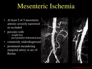
MVT - Diagnosis • CT-angiography is the imaging modality of choice • Image demonstrates portal vein thrombosis
MVT - Management • If ACUTE, then triage based on presentation • If acute abdomen, assess vitals, urgent CTA, and consider surgery if question of intestinal viability • If stable, then heparin x 7 days with Coumadin x 3-6 months • If hypercoagulable or repeat event, then consider lifelong Coumadin
MVT - Management • If CHRONIC (and asymptomatic), then endoscopy to screen for varices and do nothing • Collaterals have usually formed and taken care of the problem • Coumadinization carries more risk of bleeding than benefit at that point
NOMI • In the ER we call this “ischemic colitis” • Small arterial arcades with mini-thrombii and poor flow • Precipitated by some cardiovascular disturbance (atrial fibrillation, CHF, overmedicated on antihypertensives, sepsis, etc)
Diagnosis • Patients usually come with a history of crampy lower quadrant pain with bloody stools, on/off • Discrete episodes • Lasts hours to days • Problem usually self-limited • Medical history usually shows: over age 65, CAD, PVD, HTN, DM, lipidemia, etc.
Physical, Labs • Usually patient normal • Blood loss typically minimal, although in certain cases can be severe • DRE is mandatory • CBC, Lactic acid, renal function, electrolytes, liver enzymes and lipase • Imaging usually restricted to CT (exclude diverticulitis) only
Management • Controversy as to whether to start antibiotics • Supportive management • Early endoscopy • Biopsy • Watch for signs of deterioration over 48 hours • Optimize hemodynamics, referral to cardiology, etc