Download
1 / 48
630 likes | 2.12k Views
Assoc. Prof. Melih Güven Yeditepe University Hospital Department of Orthopaedics and Traumatology. BONE TUMORS. Learning Objectives. Classification of benign bone tumors Charasteristic clinical and radiological features of most common benign bone tumors Definition of tumor like lesions

E N D
Assoc. Prof. Melih Güven Yeditepe University Hospital Department of Orthopaedics and Traumatology BONE TUMORS
Learning Objectives • Classification of benign bone tumors • Charasteristic clinical and radiological features of most common benign bone tumors • Definition of tumor like lesions • Classification of malign bone tumors • Charasteristic clinical and radiological features of most common malign bone tumors • Charasteristic features of metastatic bone tumors • Surgical applications in benign and malign bone tumors
Bone tumors • General knowledge • Benign tumors • Tumor like lesions • Malign tumors • Metastatic bone tumors
General approaches • Initial referral • Pain • Deeply, sharp and increased over time • Localized sensitivity and rush • Pathologic fracture • Deformity • Incidental finding
General approaches • First stage evaluation • Anamnesis • Duration and level of pain, number and duration of the lesions, age, loss in weight, fever, weakness • Physical examination • Site and size of the lesion, mobility, sensitivity,LAP • Radiological evaluation • Codman triangle, onion skin view, sunrise view • Laboratory findings • ALP, serum Ca level, ESR, LDH, protein electrophoresis
Bone Tumors Imaging • Solitary or multiple lesions? • What type of bone is involved? • Which part of the bone is involved? • Are the margins of the lesion well defined? • Is there any bony reaction? • Does the lesion contain calcification?
General approaches • Second stage evaluation • CT • Bone scintigraphy – Tc 99m • Anjiography • MRI • PET – CT
General approaches • Second stage evaluation • Biopsy • Closed • Fine needle aspiration biopsy • Trochar biopsy • Needle biopsy under CT control • Open • Incisional biopsy • Excisional biopsy • Frozen section biopsy (intraoperative)
Staging • Staging of benign bone tumors (Enneking) • Stage I: Latent • Dense capsule, no reactive zone • Spontaneous healing • Stage II: Active • Dense capsule, adhesions between capsule and surrounding soft tissue; reactive zone present • No spontaneous healing
Staging • Staging of benign bone tumors (Enneking) • Stage III: Agressive • Irregularity is present at the border of the tumor, thin capsule • Dense reactive zone and edema • Tumor can grow up quickly, no restriction
Staging • Staging of malign bone tumors (Enneking) • G :G1: Low potential of metastasis , G2: High potential of metastasis • T : Intracompartmantal (T1) or extracompartmantal (T2) • M : M0: No metastasis, M1: Metastasis present IA G1 T1 M0 IB G1 T2 M0 IIA G2 T1 M0 IIB G2 T2 M0 IIIA G1-G2 T1 M1 IIIB G1-G2 T2 M1
General classification • Tumors with osseous origin a-Benign: Osteoma, Osteoid osteoma, Osteoblastoma b-Malign: Osteosarcoma • Tumors with cartilaginous origins a- Benign :Chondroma, Osteochondroma, Chondroblastoma, Chondromyxoidfibroma b- Malign : Chondrosarcoma
General classification • Tumors with bone marrow origins Ewing’s sarcoma Reticulumcellsarcoma Lymphosarcoma Multiple myeloma • Gaint cell tumors Gaint cell bone tumors (osteoclastoma)
General classification • Tumor like lesions (Fibrous, cystic) Simple bone cyst Aneurysmal Bone Cyst Fibrouscorticaldefect (Non-ossifyingfibroma) Eosinophilicgranuloma FibrousDysplasia Desmoplasticfibroma
Treatment • If possible, extremity preserving surgery • Functional extremity • External body view • Psychological copmpliance • Sociocultural compliance
Surgical treatment 1.Intracapsular excision: - Benign, latent lesions 2.Marginal excision: - Surgical borders contain also reactive zone of the tumor - Benign, active lesions
Surgical treatment 3.Wide resection: - Surgical borders contain surrounding healthy tissue - Benign, agressive lesions; malignant low grade lesions 4.Radical resection: - Tumor resection with whole compartment contents - Malignant high grade lesions 5. Amputation
Adjuvanttherapies • Chemotheraphy • Neoadjuvant (8-12 weeks) • Adjuvant (6-12 months) • Radiotheraphy • Local adjuvants: • Fenol, hydrogen peroxid, liquid nytrogene, bone cement, alcohol, coterisation, hot water (80°C)
Enchondroma • Ollier disease • Maffuci syndr.
Benign bone tumors • Osteochondroma • Multiple herediter osteochondromatisis • Enchondroma • Osteoid osteoma • Osteoblastoma • Giant cell tumor • Chondroblastoma
Fibrousdysplasia mlgn 5 yıl sonra
Tumorlikelesions • Simple bone cyst • Aneurysmal bone cyst • Eosinophilic granuloma • Fibrous cortical defect (Non-ossifying fibroma) • Fibrous dysplasia
Osteosarcoma Birkaç ay sonra
Malignant Bone Tumors • Osteosarcoma • Chondrosarcoma • Ewing’s sarcoma • Multiple myeloma • Chordoma
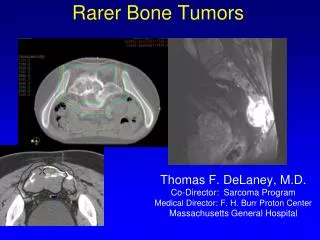
Metastatic Bone Tumors • Most common malignancy of the skeleton • Pain (most common), limitation of movement, pathologic fracture, spinal cord compression, hypercalcemia • Breast most common in woman, prostate and lung most common in man
Metastatic Bone Tumors • Spreading; • Direct • Indirect • Lenfogenous • Hematogenous • Location of metastasis; - vertebra % 69 - pelvis %41 - femur % 25 - cranium %14 Batson venous plexus
Breast met. Kidney met. Prostat met. Prostate met. ob ol Lung met.
Metastatic Bone Tumors • Diagnosis; • Bone scintigraphy (Tecnesium 99m) • Radiographs • Computarised tomography (CT) • MRI • PET-CT (F-18 PET) • Biopsy Standart methods
Metastatic Bone Tumors • Treatment • Chemotherapy • Radiotherapy • Tumor excision ? • Limb salvage surgery • Preventive surgery for pathologic fracture