Download
1 / 119
1.23k likes | 1.8k Views
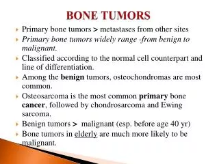
PRIMARY BONE TUMORS. DR.MUNENE. BENIGN BONE TUMOURS. The generally are Solitary Localised Slow growing Enlarges by pressure and expansion Do not metastasize (though benign tumours like GCT and chondroblastoma have been known to metastasize). GENERAL CHARACTERISTICS OF BONE TUMOURS

E N D
PRIMARY BONE TUMORS DR.MUNENE
BENIGN BONE TUMOURS • The generally are • Solitary • Localised • Slow growing • Enlarges by pressure and expansion • Do not metastasize (though benign tumours like GCT and chondroblastomahave been known to metastasize)
GENERAL CHARACTERISTICS OF BONE TUMOURS Age at presentation Peak incidence Range of common incidence 1º malignant tumours Characterized by ability to spread to other regions not in continuity Spread usually via bloodstream Also by lymph/ CSF/ serous cavities/ bone marrow spaces Unremitting growth ± periodic variation
s/s • Pain • Swelling and tenderness near the affected area • Pathological # • Constitutional S/S –Fatigue ,Unintended weight loss, Night Sweats
ROLE OF IMAGING • Detection • Diagnosis • Staging - prebiopsy - presurgery • Follow-up - mets - recurrence - complications of treatment
RADIOLOGICAL ASSESSMENT • PLAIN X-RAY • CT SCAN • RNI • MRI
mgt • Medical : Biphosphonates • Surgical : Limb Salvage Surgery : Amputation Neo- Adjuvanct Therapy : Chemo/Radiotherapy chondrosarcoma-less sensitive ewing’s sarcoma-very sensitive
RADIOLOGICAL ASSESSMENT • LOCATION • RATE OF GROWTH • REGULARITY OF THE MARGIN • PERIOSTEAL REACTION • MATRIX MINERALIZATION
RADIOLOGICAL ASSESSMENT • LOCATION • Within the skeleton • Within individual bone • RATE OF GROWTH • The radiologist fairs better than the histopathologist • Advancing margin of the lesion: in an actively growing tumour there is little peripheral screlosis. Conversly, least aggressive lesions may be surrounded by a rim of sclerosis
RADIOLOGICAL ASSESSMENT • REGULARITY OF THE MARGIN • Regular margin and endosteal destruction of the cortex signifies slow growth • PERIOSTEAL REACTION • There are various types, none being pathognomonic of any particular tumour • It helps to indicate aggressiveness of the tumour
RADIOLOGICAL ASSESSMENT • MATRIX MINERALIZATION • This depends on extracellular material produced by the tumour: • Chondral Ca+ are typically linear, curvilinear, ring-like, punctate or nodular • Osseous mineralization is clowd-like & poorly defined • Fibrous tumours produce the characteristic ‘ground-glass’ appearance
PERIOSTEAL REACTION • The periosteum is a membrane several cell layers thick that covers almost all of every bone • The only parts not covered by this membrane are covered by cartilage • Besides covering the bone and sharing some of its blood supply with the bone, it also produces bone when it is stimulated appropriately
PERIOSTEAL REACTION • With slow-growing lesions, the periosteum has time to produce new bone • With rapidly growing lesions, the periosteum cannot produce new bone as fast. An interrupted pattern results, which may be: • a thin shell of calcified new bone • one or more concentric shells of new bone over the lesion, sometimes called lamellated or "onion-skin" periosteal reaction.
PERIOSTEAL REACTION • If the lesion grows rapidly but steadily, the periosteum will not have enough time to lay down even a thin shell of bone • In such cases, the tiny fibers that connect the periosteum to the bone (Sharpey'sfibers) become stretched out perpendicular to the bone. When these fibers ossify, they produce a pattern sometimes called "sunburst" or "hair-on-end" periosteal reaction, depending of how much of the bone is involved by the process.
BENIGN BONE TUMOURS • CHONDROID ORIGIN • OSTEOID ORIGIN • CYSTS • FIBROUS ORIGIN
CHONDROID ORIGIN • Chondroma • Osteochondroma • Chondroblastoma • Chondromyxoid fibroma
CHONDROMA • Commonly central in situation, whence it is referred to as enchondroma, but may be eccentric e.gsubperiosteal location – periostealchondroma • Multiple enchondromas are found Ollier’sdse & Maffucci’s syndrome • Affect tubular bones of the hands & feet in >50%. • If it becomes painful in absence of fracture, it should be biopsied.
CHONDROMA • Age range of 10-80 yrs, most being in 2nd to 4th decades • 40% occur in the hands, being x5 more common here than in the feet • Are extremely rare in the spine and almost unknown in the skull
CHONDROMA, radiology • Most arise in the medulla of the phalanges • Often eccentric and 75% are solitary • Well circumscribed, round lytic lesions which expand the cortex • Flecks of Ca+ may be seen within
CHONDROMA, radiology • Features of benignity include: • <20 yrs • Purely medullary with well formed Calcification • Well-defined round margin • Cortex intact • Slow growth
CHONDROMA, rare types Multiple chondromas (Ollier’s disease, enchondromatosis) • Not genetically inherited • Predominatly unilateral, in respect to the side of the body and location within each bone • Malignant change uncommon
CHONDROMA, rare types • Enchondromatosis with haemangiomas (Maffucci’s syndrome) • Incidence of chondrosarcomatous change in uncertain due to rarity
OSTEOCHONDROMA • Cartilage-capped exostosis • It is the cartilage cap that is actively growing • Is a developmental anomaly • Radiotherapy in childhood reported to cause it • Peaks in the 2nd decade, but can occur from 2 to 60 yrs • M:F ratio is 1.4:1
OSTEOCHONDROMA • Long bones commonly affected, esp around the knee • The lesions are metaphyseal but migrate to the diaphysis as normal metaphyseal growth occurs • This gives ‘coat-hanger’ variety, pointing away from adjacent joint • May be single or several
OSTEOCHONDROMA • Multiple cartilaginous exostoses (diaphyseal aclasis) are inherited in an AD fashion • They may be larger than the solitary variety & may lead to shortening or deformity of the affected limbs • MRI is highly accurate in the assessment of symptomatic osteochondromas
OSTEOCHONDROMA • Presentation tends to relate to mechanical problems, an enlarging mass, pressure on adjoining structures, or occasionally fracture • As the lesion grows, the marrow cavity extends into the exostosis • Actively growing cartilage is not visible radiologically, but shown on US, CT or MRI • Become Ca+ with age in a punctate nodular fashion
OSTEOCHONDROMA • Small exostoses (not cartilage-capped) may follow trauma • These tend to remain static or regress • Complete resection of a solitary osteochondroma cures the patient • Malignant transformation of solitary osteochondroma is rare. When it occurs, it is usually chondrosarcoma, although a rare osteosarcoma has been reported
CHONDROBLASTOMA • Rare, usually presenting in a child with pain in the region of a joint • Typically found in epiphysis of a long bone • Though generally benign, cases of localized aggressive spread associated with distant mets reported • 80% occur in the second decade • M:F ratio is 2:1
CHONDROBLASTOMA • Usually eccentric in the epiphyses but may straddle the growth plate if closure is partial
CHONDROBLASTOMA • The plain film is usually sufficient for diagnosis. • Treatment is curettage, but there is a 15% recurrence rate. • To decrease this recurrence rate, CT is used to further evaluate extent of the lesion. • In this case, there is in fact a small finger of the lesion extending anteriorly into the lateral portion of the tibial epiphysis
CHONDROBLASTOMA, radiology • Is spherical & well defined • Fine sclerotic margin • May be destructive thereby suggesting malignancy • Central punctate Ca+ seen in 10% on plain films • Secondary ABC change may be confirmed by fluid-fluid levels
CHONDROBLASTOMA, differential • Sub-chondralcyst • GCT, which are rare in children. When they occur, they tend to be metaphyseal • Chondrosarcoma, clear-cell variety • Brodies abscess considered if involves both epiphysis and metaphysis, including simple & ABC
CHONDROMYXOID FIBROMA • Accounts for <0.5% of biopsied 1º bone tumors • Presentation is with local pain & tenderness • Lesion consists of chondroid, myxoid & fibrous elements in almost any proportion • 75% of cases seen btn10-30 yrs, peaking in the 2nd decade • There is a slight male preponderance
CHONDROMYXOID FIBROMA • Most lesions are metaphyseal and eccentric within the medulla • It results in thinning and expansion of the cortex • Prox 1/3 of tibia contributes to 25% of all cases
CHONDROMYXOID FIBROMA, radiology • Margins are well demarcated and lobulated with focal histological Ca+ in a 1/3 of cases • Involves cortex & medulla • A sclerotic endosteal border is characteristic • Demonstrable Ca+ only in 13%
CHONDROMYXOID FIBROMA, radiology • It may show a trabeculated or ‘soap-bubble’ appearance due to endosteal ridging • MRI does not offer information to distinguish it from other tumours • Recurrence of 25% following curretage • Malignant transformation virtually unknown
CHONDROMYXOID FIBROMA, differential • Outside its classical location, less typical features may resemble those of other benign tumours: • ABC • Enchondroma • Chondroblastoma • Chondrosarcoma • Fibrous dysplasia
CYSTS OF BONE • SIMPLE BONE CYST (unicameral cyst) • ABC • GCT
SIMPLE BONE CYST • Unicameral cyst • Perhaps the only true primary cyst of bone • Aet is unknown • Usually solitary • 70% btwn 4-10 yrs with 90% being <20 yrs
SIMPLE BONE CYST • M:F ratio is 2:1 • Rarely symptomatic • Pathological fracture is common in pre-adolescent, esp when the humerus is involved • The prox 1/3 humerus is the commonest site then the prox 1/3. This gives 80% btwn the two
SIMPLE BONE CYST • Other sites of predilection iclude the calcaneous & ilium, which tend to be affected in the older age range • Treatment is by steroid injection, observation of minimally symptomatic lesions, or by curettage and bone grafting
ANEURYSMAL BONE CYST • The concept that many ABCs are secondary to preceding benign or malignant lesions is now generally accepted • Most pts in the 2nd decade • Sex incidence is equal
ANEURYSMAL BONE CYST • Majority present with pain & local swelling • Spinal lesions may result in scoliosis or neurological symptoms • At surgery, the cyst-like lesion is found. Curretage mayresult in brisk bleeding • Any site is affected
GIANT CELL TUMOUR • Is the 5th commonest primary tumour of bone • Its behaviour in unpredictable: most lesions are locally aggressive while a few are overtly malignant & metastasize • Local recurrence after curretage may occur in up to 50%
GIANT CELL TUMOUR • In GCT, the smaller stromal cells are actually the tumourous cells (which are thought to be derived from osteoblasts) • 80% are in pts btwn 18-45yrs, rare before skeletal maturity • M:F ratio 2:3 • Pathological fracture may be found in 10%
GIANT CELL TUMOUR • About 50% are found on either side of the knee joint, distal end of femur being the commonest single site • When in an immature pt, it involves the metaphysis & may rarely cross the growth plate • In the spine, the sacrum is most commonly affected, to lie adjacent to the sacro-iliac joint
GIANT CELL TUMOUR • The lesion is highly vascular, but forms neither osteoid nor cartilage • Occasional blood-filled spaces mimic ABC under the microscope. • Tumours with giant cell will be part of the histopathologist ddx, and include: • Chondroblastoma, • Brown tumour of hyperparathyroidism • ABC • Osteoid-rich osteosarcoma • Benign fibrous histiocytoma
FIBROUS ORIGIN • FIBROUS CORTICAL DEFECT (metaphyseal fibrous defect) • NONOSSIFYING FIBROMA (fibroxanthoma) • BENIGN FRBROUS HISTIOCYTOMA • DESMOPLASTIC FIBROMA • POST-TRAUMATIC CORTICAL DESMOID