LUNG
LUNG. Trachea divides into right and left mainstem bronchi Each main bronchus divides into lobar bronchi, then into segmental bronchi Lobar bronchi are usually called secondary bronchi and segmental bronchi are called tertiary bronchi Bronchioles lack cartilage and submucosal glands

LUNG
E N D
Presentation Transcript
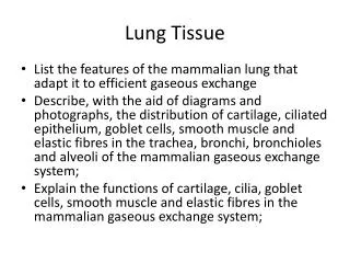
Trachea divides into right and left mainstem bronchi • Each main bronchus divides into lobar bronchi, then into segmental bronchi • Lobar bronchi are usually called secondary bronchi and segmental bronchi are called tertiary bronchi • Bronchioles lack cartilage and submucosal glands • Right lung has 3 lobes, left lung has 2 lobes plus lingula • Right bronchus more vertical than left, thus aspirated material tends to enter right lung • Lung has double arterial supply - pulmonary and bronchial
Further branching of bronchioles leads to the terminal bronchioles, which are less than 2 mm in diameter. • The part of the lung distal to the terminal bronchiole is called the acinus; • It is approximately spherical, with a diameter of about 7 mm. An acinus is composed of respiratory bronchioles (emanating from the terminal bronchiole), which give off several alveoli from their sides. These bronchioles then proceed into the alveolar ducts, which immediately branch into alveolar sacs, the blind ends of the respiratory passages, whose walls are formed entirely of alveoli, which are the site of gas exchange. • A cluster of three to five terminal bronchioles, each with its appended acinus, is usually referred to as the pulmonary lobule.
Alveoli are lined by respiratory epithelium (pseudostratified, columnar, ciliated) • Alveoli contain type I and II pneumocytes • Type I pneumocytes: 95%, flattened • Type II pneumocytes: 5%, produce surfactant (lamellar bodies on EM), involved in repair if type I destroyed
The microscopic structure of the alveolar walls (or alveolar septa) consists, from blood to air, of the following • The capillary endothelium lining the intertwining network of anastomosing capillaries. • A basement membrane and surrounding interstitial tissue separating the endothelial cells from the alveolar lining epithelial cells. • Alveolar epithelium, which contains a continuous layer of two principal cell types: flattened • Alveolar macrophages, loosely attached to the epithelial cells or lying free within the alveolar spaces, derived from blood monocytes and belonging to the mononuclear phagocyte system. Often, they are filled with carbon particles and other phagocytosed materials.
ACUTE RESPIRATORY DISTRESS SYNDROME (DIFFUSE ALVEOLAR DAMAGE) Acute respiratory distress syndrome (ARDS) (synonyms include "shock lung," "diffuse alveolar damage," "acute alveolar injury," and "acute lung injury") is a clinical syndrome caused by diffuse alveolar capillary damage. It is characterized clinically by the rapid onset of severe life-threatening respiratory insufficiency, cyanosis, and severe arterial hypoxemia that is refractory to oxygen therapy and that may progress to extrapulmonary multisystem organ failure.
Obstructive Versus Restrictive Pulmonary • (1) obstructive disease (or airway disease), characterized by an increase in resistance to airflow owing to partial or complete obstruction at any level, from the trachea and larger bronchi to the terminal and respiratory bronchioles, and • (2) restrictive disease, characterized by reduced expansion of lung parenchyma, with decreased total lung capacity. • Although many conditions have both obstructive and restrictive components, distinction between the two patterns of pulmonary dysfunction is useful in correlating the results of pulmonary function tests with the radiologic and histologic findings in individual patients
The major diffuse obstructive disorders are emphysema, chronic bronchitis, bronchiectasis, and asthma • Emphysema and chronic bronchitis are often clinically grouped together and referred to as chronic obstructive pulmonary disease (COPD), since many patients have overlapping features of damage at both the acinar level (emphysema) and bronchial level (bronchitis), almost certainly because one extrinsic trigger-cigarette smoking-is common to both. In most patients, COPD is the result of long-term heavy cigarette smoking; about 10% of patients are nonsmokers
Chronic bronchitis :persistent cough with sputum for 3 months in 2 consecutive years • More infections, purulent sputum, hypercapnia, hypoxia than emphysema; clinically called “blue bloaters” • May cause secondary pulmonary vascular hypertension, corpulmonale, congestive heart failure; death due to respiratory acidosis and coma, congestive heart failure, pneumothorax • Causes: 4-10x more common in smokers, also chronic irritation, infections • Tobacco interferes with ciliary action, directly damages airway epithelium, inhibits ability of white blood cells to clear bacteria; infections maintain but do not initiate chronic bronchiti • Micro:early-hypersecretion of mucus in large airways with hypertrophy of submucosal glands in tracheobronchial tree • later-increase in goblet cells in small airways causes excessive mucus production and airway obstruction
Emphysema is a condition of the lung characterized by abnormal permanent enlargement of the airspaces distal to the terminal bronchiole, accompanied by destruction of their walls and without obvious fibrosis. In contrast, the enlargement of airspaces unaccompanied by destruction is termed "overinflation," for example, the distention of airspaces that occurs in the remaining lung after unilateral pneumonectomy.
Emphysema is classified according to its anatomic distribution within the lobule. Recall that the lobule is a cluster of acini, the alveolated terminal respiratory units. Although the term "emphysema" is sometimes loosely applied to diverse conditions, • (1) centriacinar, (2) panacinar, (3) paraseptal, and (4) irregular. • The first two cause clinically significant airflow obstruction Centriacinar emphysema is far more common than the panacinar form, constituting more than 95% of cases. Clinical management does not rely on precise anatomic diagnosis and classification, which, however, do provide important clues to pathogenesis.
centriacinar (Centrilobular) Emphysema. The distinctive feature of this type of emphysema is the pattern of involvement of the lobules; the central or proximal parts of the acini, formed by respiratory bronchioles, are affected, whereas distal alveoli are spared • Thus, both emphysematous and normal airspaces exist within the same acinus and lobule. The lesions are more common and usually more severe in the upper lobes, particularly in the apical segments • Centriacinar emphysema occurs predominantly in heavy smokers, often in association with chronic bronchitis.
Panacinar (Panlobular) Emphysema. In this type, the acini are uniformly enlarged from the level of the respiratory bronchiole to the terminal blind alveoli .The prefix "pan" refers to the entire acinus but not to the entire lung. In contrast to centriacinar emphysema, panacinar emphysema tends to occur more commonly in the lower zones and in the anterior margins of the lung, and it is usually most severe at the bases. This type of emphysema is associated with α1-antitrypsin (α1-AT) deficiency. • Distal Acinar (Paraseptal) Emphysema. In this type, the proximal portion of the acinus is normal, but the distal part is predominantly involved. The emphysema is more striking adjacent to the pleura, along the lobular connective tissue septa, • Airspace Enlargement with Fibrosis (Irregular Emphysema).Irregular emphysema, so named because the acinus is irregularly involved, is almost invariably associated with scarring.and at the margins of the lobule
This is a more subtle appearance for centrilobular emphysema in which there are "dirty holes" that appear focally where the central portions of lung acini have lost lung parenchyma while collecting anthracotic pigment at the same time. This pattern is typical for smokers.
Bullousemphysema: any form that produces blebs > 1 cm; often subpleural, near apex, associated with tuberculosis scarring; may rupture and cause pneumothorax, hemorrhage; called placental transmogrification if it resembles chorionic villi • Interstitial emphysema: air into connective tissue stroma of lung, mediastinum or subcutaneous tissue; due to alveolar tears, chest wounds, coughing, whooping cough
Asthma :Chronic relapsing inflammatory disorder characterized by hyperreactive airways, causing episodic, reversible bronchoconstriction • Usually associated with atopy, mediated by IgE • Extrinsic: Type I hypersensitivity; either atopic (due to allergens), occupational or due to allergic bronchopulmonaryaspergillosis • Intrinsic:nonimmune; due to aspirin ingestion, pneumonia, cold, stress, exercise
Gross:overdistended lungs, small areas of atelectasis, thick mucus plugs in proximal bronchi containing whorls of shed epithelium • Micro:eosinophilscontain Charcot-Leyden crystals (eosinophil membrane protein); increased mucosal goblet cells and submucosal glands, thickened basement membrane, bronchial smooth muscle hypertrophy, airway wall edema
BRONCHIECTASIS is a disease characterized by permanent dilation of bronchi and bronchioles caused by destruction of the muscle and elastic tissue, resulting from or associated with chronic necrotizing infections. To be considered bronchiectasis, the dilation should be permanent; reversible bronchial dilation often accompanies viral and bacterial pneumonia • Bronchiectasis develops in association with a variety of conditions, which include the following:3Congenital or hereditary conditions, including cystic fibrosisintralobar sequestration of the lung (discussed earlier in this chapter), immunodeficiency states, and primary ciliarydyskinesia and Kartagener syndromes (discussed later) • Postinfectious conditions, including necrotizing pneumonia caused by bacteria (Mycobacterium tuberculosis, Staphylococcus aureus, Haemophilusinfluenzae, Pseudomonas), viruses (adenovirus, influenza virus, HIV), and fungi (Aspergillus species) • Bronchial obstruction, owing to tumor, foreign bodies, and occasionally mucus impaction, in which the bronchiectasis is localized to the obstructed lung segment • Other conditions, including rheumatoid arthritis, systemic lupus erythematosus, inflammatory bowel disease, and post-transplantation (chronic lung rejection, and chronic graft-versus-host disease after bone marrow transplantation)
Bronchiectasis usually affects the lower lobes bilaterally, particularly air passages that are vertical, and is most severe in the more distal bronchi and bronchioles. When tumors or aspiration of foreign bodies leads to bronchiectasis, the involvement may be sharply localized to a single segment of the lung. The airways are dilated,sometimes up to four times normal size. These dilations may produce long, tubelike enlargements (cylindrical bronchiectasis) or, in other cases, may cause fusiform or even sharply saccular distention (saccularbronchiectasis).
Characteristically, the bronchi and bronchioles are sufficiently dilated that they can be followed, on gross examination, directly out to the pleural surfaces. • By contrast, in the normal lung, the bronchioles cannot be followed by ordinary gross dissection beyond a point 2 to 3 cm removed from the pleural surfaces. On the cut surface of the lung, the transected dilated bronchi appear as cysts filled with mucopurulent secretions
The histologic findings vary with the activity and chronicity of the disease. • In the full-blown, active case, there is an intense acute and chronic inflammatory exudation within the walls of the bronchi and bronchioles, associated with desquamation of the lining epithelium and extensive areas of necrotizing ulceration. • There may be pseudostratification of the columnar cells or squamousmetaplasia of the remaining epithelium. In some instances, the necrosis completely destroys the bronchial or bronchiolar walls and forms a lung abscess.
Fibrosis of the bronchial and bronchiolar walls and peribronchiolar fibrosis develop in the more chronic cases, leading to varying degrees of subtotal or total obliteration of bronchiolar lumina • In the usual case of bronchiectasis, a mixed flora can be cultured from the involved bronchi, including staphylococci, streptococci, pneumococci, enteric organisms, anaerobic and microaerophilic bacteria, and (particularly in children) Haemophilusinfluenzae and Pseudomonas aeruginosa. In
The mid lower portion of this photomicrograph demonstrates a dilated bronchus in which the mucosa and wall is not clearly seen because of the necrotizing inflammation with destruction.
Pneumonia • viral pneumonia predisposes to bacterial pneumonia • Common agents: Staphylococcus aureus, Streptococcus pneumoniae, Haemophilus influenza, Pseudomonas aeruginosa, coliforms • Complications: abscess, empyema, organization, sepsis, meningitis • Consolidation:exudative solidification of lung
Bacterial pneumonia has two gross patterns of anatomic distribution: lobular bronchopneumonia and lobar • Patchy consolidation of the lung is the dominant characteristic of bronchopneumonia . • Lobar pneumonia is an acute bacterial infection resulting in fibrinosuppurative consolidation of a large portion of a lobe or of an entire lobe These anatomic but still classic categorizations are often difficult to apply in the individual case because patterns overlap.
Foci of bronchopneumonia are consolidated areas of acute suppurative inflammation. The consolidation may be patchy through one lobe but is more often multilobar and frequently bilateral and basal because of the tendency of secretions to gravitate into the lower lobes. Well-developed lesions are usually 3 to 4 cm in diameter, slightly elevated, dry, granular, gray-red to yellow, and poorly delimited at their margins • Histologically, the reaction usually elicits a suppurative, neutrophil-rich exudate that fills the bronchi, bronchioles, and adjacent alveolar spaces.
More virulent bacteria and/or more severe pneumonias can be associated with destruction of lung tissue and hemorrhage. Here, alveolar walls are no longer visible because there is early abscess formation. There is also hemorrhage.
Complications of pneumonia include (1) tissue destruction and necrosis, causing abscess formation (particularly common with type 3 pneumococci or Klebsiella infections); (2) spread of infection to the pleural cavity, causing the intrapleuralfibrinosuppurative reaction known as empyema; (3) organization of the exudate, which may convert a portion of the lung into solid tissue and (4) bacteremic dissemination to the heart valves, pericardium, brain, kidneys, spleen, or joints, causing metastatic abscesses, endocarditis, meningitis, or suppurative arthritis.
CARCINOMAS • Lung cancer is currently the most frequently diagnosed major cancer in the world and the most common cause of cancer mortality worldwide. • Squamous cell carcinoma (25% to 40%) • Adenocarcinoma (25% to 40%) • Small cell carcinoma (20% to 25%) • Large cell carcinoma (10% to 15%)