Download
1 / 26
360 likes | 831 Views
The Motor System. Chapter 8. Overview. The motor system is divided into 4 components: Direct activation pathway Control circuits Indirect activation pathway Final common pathway. Direct activation pathway Largest, best-defined motor pathway

E N D
The Motor System Chapter 8
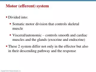
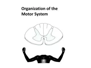
Overview • The motor system is divided into 4 components: • Direct activation pathway • Control circuits • Indirect activation pathway • Final common pathway
Direct activation pathway • Largest, best-defined motor pathway • It is a “single neuron” pathway, extending from the cerebral cortex to the spinal cord (corticospinal tract) • Major function: to effect voluntary activity, particularly skilled movements under conscious control
Direct activation pathway • Origins: • Primary motor cortex • Premotor cortex • Supplementary motor cortex • Major neurotransmitters: glutamate (excitatory) and GABA (inhibitory) • Damage results in weakness, with loss of voluntary movements especially fine, skilled movements, but preservation of other forms of movements including segments reflexes
Control Circuits • 2 parallel pathways, the cerebellar and basal ganglia, control and modify motor activity • They both receive input from several motor and sensory cortical areas and project back to the cerebral cortex via the thalamus. They integrate and modulate motor activity primarily through the cerebral cortex and direct activation pathways.
Control Circuits • They also send information to the brainstem and the indirect activation pathways. • Basal ganglia • Concerned with learned, automatic behavior and with preparing and maintaining the background support, or posture, needed for voluntary motor activity. (Neurotransmitters: dopamine and GABA) • Cerebellum • Accomplishes the coordination and correction of movement errors of muscles during active movements. (Neurotransmitters: glutamate and GABA)
Indirect activation pathways • The older, more diffuse motor pathways, sometimes called extrapyramidal pathways • These pathways mediate the enormous number of automatic activities involved in normal motor function. For example, the maintenance of erect posture when sitting or standing requires the coordinated contraction of many muscles.
Indirect activation pathways • This coordination is under subconscious control and is mediated by the indirect activation pathways which are: • Reticulospinal • Vestibulospinal • Rubrospinal • Diseases affecting these pathways are manifested with abnormal muscle tone and reflexes.
Final common pathway • The somatic (skeletal) muscles that perform the voluntary movements are all under direct control of lower motor neurons and contract only in response to activation by these neurons. • These neurons are located in the ventral horn of the spinal cord and brainstem. • A lower motor neuron and the muscle fibers under its control constitute a motor unit. • The final common pathway consists of many motor units through which all activities in the motor system must act. • Neurotransmitter: acetylcholine
Internal capsule Corticobulbar fibers occupy a more anterior location in the posterior limb than the corticospinal
Clinical Correlations Knowledge of the clinical manifestations of the diseases affecting the direct activation pathway comes from experimental studies and from observation of clinical disorders. Disturbances of the corticospinal system may be irritative (positive) or paralytic (negative).
Clinical Correlations: irritative vs. paralytic Irritative (seizures) Jacksonian seizures – focal motor seizures starting from the distal upper extremity spreading proximally Paralytic Weakness – fine movements, skilled movements (distal muscles); no atrophy; presence of Babinski sign and primitive reflexes (upper motor neuron manifestations)
Control Circuits BASAL GANGLIA learned, automatic behavior preparing and maintaining the background support or posture needed for voluntary motor activity Anatomy and Physiology caudate nucleus, lentiform nucleus (putamen, globus pallidus), substantia nigra, subthalamic nucleus striatum=caudate nucleus + putamen striatum neurotransmitters: acetylcholine, dopamine (produced in the substantia nigra and transported to striatum via the nigrostriatal pathway)
Control Circuits: Basal Ganglia Clinical Correlations: excess discharge slowing (hypokinesia) lack of discharge hyperactivity (involuntary movements) Involuntary movements athetosis chorea tremors dystonia ballismus
Control Circuits: Basal Ganglia Clinical Correlation: Speech hypokinetic – quiet, weak, monotonous voice, poor articulation, short rushes of words, breathy quality hyperkinetic – bursts of loudness, elevation of pitch, voice stoppages, distortion of vowels, disintegration of articulation
Control Circuits CEREBELLUM coordination and correction of movement errors of muscles during active movements neurotransmitters: glutamate, GABA
Control Circuits: Cerebellum Functional subdivisions Vestibulocerebellum Flocculonodular lobe connected with the vestibular nuclei through the inferior cerebellar peduncle Controls and coordinates axial musculature and movements of the head and eyes
Control Circuits: Cerebellum Functional subdivisions Spinocerebellum Made up of vermis and paravermis Its primary input is from dorsal spinocerebellar tract (via the inferior cerebellar peduncle) and the ventral spinocerebellar tract (via the superior cerebellar peduncle) which provide information about motor performance and motor neuron excitability, respectively.
Control Circuits: Cerebellum Functional subdivisions Cerebrocerebellum Made up of the lateral portions of the cerebellar hemispheres and receives inputs from pontine nuclei via the middle cerebellar peduncle The output is from the dentate nucleus through the superior cerebellar peduncle This subdivision is important in initiation of movement and in coordination of muscles (specifying direction, pattern, and intensity of movement)
Cerebellum: Clinical Correlations • Flocculonodular Syndrome • Occurs with midline tumors in children • Produces truncal ataxia, with unsteadiness and falling and disturbances with gait and standing; with nystagmus
Cerebellum: Clinical Correlations • Anterior Lobe Syndrome • Commonly due to degenerative diseases of the adults • Produces severe gait ataxia (broad-based) without ataxia of the upper limbs, and mild ataxia of the legs • Cerebellar Hemisphere Disease • Produces limb ataxia with hypotonia, intention tremors, dyssynegria and dysmetria ipsilateral to the lesion
Cerebellum: Clinical Correlations • Disorders of speech also occur in cerebellar disease, particularly when it involves the vermis. • These disorders are manifested as irregularities of pitch, loudness, and rhythm and are known as ataxic dysarthria. • Many cerebellar diseases can be reversed if they are identified early and treated appropriately. • Cerebellar degeneration produced by vitamin E deficiency • Small cell lung carcinoma and ovarian cancers produce antibodies to Purkinje cells than cause cerebellar degeneration.
Final Common Pathway Motor unit – a lower motor neuron (ventral horn cell/CN nucleus) + muscle fibers under its control alpha motor neuron axon (nerve) synapse muscle fibers Final common pathway – many motor units through which all activities in the motor system must act