The Pulp
The Pulp. Dr Jamal Naim PhD in Orthodontics. The Pulp. It is a specialized loose connective tissue occupying a cavity in the dentine. It is a remnant of the embryologic organ for tooth development. It has an ectomesenchymal origin. The Pulp.

The Pulp
E N D
Presentation Transcript
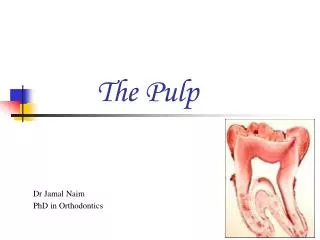
The Pulp Dr Jamal Naim PhD in Orthodontics
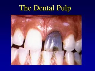
The Pulp It is a specialized loose connective tissue occupying a cavity in the dentine. It is a remnant of the embryologic organ for tooth development. It has an ectomesenchymal origin.
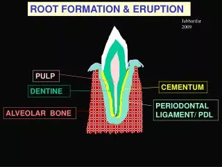
The Pulp At first the pulp is called dental papilla; which is highly vascular; and its cells develop into fibroblasts and odontoblasts. After dentin formation this tissue is called dental pulp. dental papilla
pulp dental papilla Both dentin and pulp have a common origin from the dental papilla.
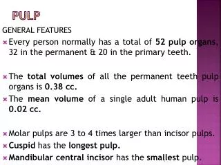
Anatomy of Pulp Pulp horns or cornua Pulp Chamber or coronal pulp, located in the crown of the tooth. Root canal or radicular pulp, is the portion of the pulp located in the root area. The apical foramen is the opening from the pulp at the apex of the tooth to be continuous with the periapical tissue . Accessory canals or lateral canal, extra canal located on the lateral portions of the root.
The coronal pulp It is called pulp chamber, where in young teeth it is large and have the shape of the D.E.J. The pulp chamber has pulp horn (s) according to the number of the cusps. Pulp horns
The radicular pulp • In anterior teeth it is single, in posterior teeth it is multiple. • Its shape conforms to the D.C.J. • Apically the root canal opens to the periodontal ligament by apical foramen. Pulp horns Root canals
The apical foramen • The pulp organs are continuous with the periapical tissue through the apical foramen. • The average size of the apical foramen is: • maxillary teeth: 0,4 mm • mandibular teeth: 0,3 mm apical foramen
The apical foramen As the root begins to develop, the apical foramen is actually larger than the pulp chamber, but it becomes more constricted at the completion of root formation.
Accessory canals Acc. canal The pulp cavity is sometimes connected with the periodontal tissue with an opening rather than the apical foramen, the lateral, accessory or supplementary canal. pulp PDL
Accessory canals They are numerous in the apical third of the root and in the bifurcation of multirooted teeth. If the root canal breaks up into multiple tiny canals, it is referred to as a delta system because of its complexity.
Etiology of accessory canals • Degeneration of the epithelial root sheath of Hertwig before odontoblasts differentiation. • Large blood vessel disturbs the course of the root sheath. • Lack of union of the tongue like projection (in multi-rooted teeth).
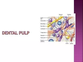
Microscopic Zones in Pulp (peripheral zone) (Weil zone) (central zone)
Microscopic Zones in Pulp (peripheral zone) (Weil zone) (central zone)
Odontoblastic process Predentin Cell bodies Odontoblasts Cell-free zone Cell-rich zone
Odontoblastic layer Dentin
Odontoblastic layer • Adjacent to predentin • Odontoblast bodies (perikaryon) in the pulp and processes (tomes fibre) in dentin. Odontoblastic layer
Odontoblastic layer Tomes process Cell body
Cell free zone (Weil zone) Dentin
Cell free zone (Weil zone) • It is about 40 micron thick. • It is prominent in the coronal pulp and less prominent in the radicular pulp. Cell free zone • It contains few cells. • In the zone many processes of the fibroblasts of the cell rich zone, nerves and blood vessels are observed.
Cell free zone (Weil zone) Odobtoblasts Blood vessel Nerves
Cell rich zone Dentin
Cell rich zone It is present between the cell free zone and the pulpal core It is composed of: • Fibroblasts, • undifferentiated mesenchymal cells • extensive vascular system Cell rich zone
Cell rich zone Odobtoblasts Fibroblasts Nerves
Pulpal core Dentin
Cross-section from the central pulp showing major support systems, including arterioles (A) with a muscular wall, thin-walled lymphatics (L), venules (V), and nerve bundles (NB)
Cells of the pulp • Progenitor cells (UMC) • Synthetic cells • Odontoblasts • Fibroblasts • Defensive cells • Histocytes • Lymphocytes • Mast cells • Plasma cells
Progenitor cells (UMC) They are also called Reserve Cells. They are descendants of undifferentiated cells in the primitive dental papilla. They are multipotential cells and likely a fibroblast type. They are found along the blood vessels They retain the capability of dedifferentiating on demand into many of the mature cell types.
Odontoblasts They line the pulpal surface of dentin A layer of predentin is always present between them and dentin. Each odontoblast has a cell body (perikaryon) and a protoplasmic process (tomes fiber) tomes fiber predentin perikaryon
Odontoblasts The odontoblasts are arranged in a single layer where: • In the pulp chamber the cells are crowded and appear psudostratefied columnar. • Near the beginning of the root canal they become columnar in shape.
Odontoblasts • In the mid portion of the root canal they become cuboidal in shape. • In the apical area of the root canal they become flat cells.
Fibroblasts They are star like cells with ovoid nucleus. The greatest number in the dental pulp, particularly in the cell rich zone. Their function is to form and maintain the pulp matrix. They also eliminate excess collagen
Defensive cells Histocytes and macrophages : • Large oval or spindle shaped • The cytoplasm is granular, especially during inflammation • Have large lysosomes • Main function is to eliminate dead cells.
Defensive cells Lymphocytes and plasma cells: • These cells are not normally present in healthy pulp tissue but are associated with injury and resultant immune responses attempts to destroy, damage, or neutralize foreign substance. • These inflammatory cell types generally appear following invasion into the area of injury.
Defensive cells • Their presence would therefore indicate the presence of a persistent irritant. plasma cells Lymphocyte
Defensive cells • Plasma cells produce antibodies. • The nucleus is eccentric with cart wheel appearance. plasma cell
Defensive cells Mast cells: • Mast cells are seldom in large numbers in normal, healthy pulps, but are commonly found in inflamed pulps. • Have granular cytoplasm with round nucleus.
Defensive cells • The granules of these cells contain histamine, a potent inflammatory mediator, and heparin. • These cells release these granules or degranulate into the surrounding tissue fluid during inflammation.
Matrix and ground substance • The extracellular compartment (matrix) is a structureless mass, gel-like in consistency and consists of collagen fibers and ground substance. • The ground substances resembles that of other loose connective tissue. • It consists of acid mucopolysaccharides, neutral glycoprotein and water. • It acts as a medium for the transport of nutrients.
Matrix and ground substance • These fibers form a loose, reticular network to support other structural elements of the pulp. • Collagen is synthesized and secreted by odontoblasts and fibroblasts. • However, the type of collagen secreted by odontoblasts to subsequently mineralize differs from the collagen produced by pulpal fibroblasts, which normally does not calcify.
Matrix and ground substance • The collagen fibers are particularly type I and type III collagen. • In young pulps there are single fibers scattered between the cells. As age change the pulp become fibrilar. • The greatest concentration of collagen fibers occurs in the most apical portion of the pulp.
Matrix and ground substance • The bundles of collagen at the pulp periphery are termed von Korff’s fibers. • They are corkscrew-like and originating between odontoblasts to pass into the dentin matrix.