Ophthalmology Case Study: Uveitis Diagnosis in a Complex Medical History
Follow a 58-year-old male with multiple health conditions and recurrent uveitis episodes seeking treatment for eye pain, blurred vision, and redness. Gain insights into diagnostic challenges and treatment strategies.


Ophthalmology Case Study: Uveitis Diagnosis in a Complex Medical History
E N D
Presentation Transcript
Case HBB Juan G. Santiago, MD Department of Ophthalmology University of Puerto Rico
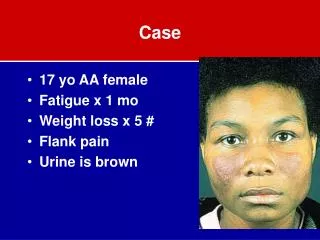
Chief Complaint • “Dolor, enrojecimiento e hinchazon del ojo derecho”
History of Present Illness • HBB is a 58 y/o male patient with hx of HTN, Multiple Sclerosis (dx 10 years ago), medical hx of syphilis, illicit drugs abuse, migraines, multiple episodes of uveitis that presents with a complaint of right eye pain and blurred vision that had worsened during a period of 2 weeks. • On Nov/07 right eye anterior uveitis was dx, with (+)RPR titers. Patient received 14 days of Penicillin G IV with improvement of condition.
History of Present Illness • On Jan/08, patient was found with right eye anterior uveitis and was send to ER to r/o syphilitic uveitis and new workup. Patient was going to be admitted but he refused, workup was not performed. Patient received home IV therapy with Ceftriaxone 2gm daily, PF oph and atropine oph. Patient refers improvement of symptoms with treatment, reason for which he did not look further medical care.
History of Present Illness • On Feb/08, patient refers right eye pain, blurred vision associated with eye redness, headaches and lid swelling that had worsened for 2 weeks. Patient refers (+) lacrimation, (-) purulent secretions, (+) photophobia, (-) flashlights, (-) curtain effect, (-) floaters. Patient denies recent trauma, viral illness, insect bite or any associated systemic symptoms.
Past Medical History • Medications • Antihypertensives (Amlodipine, HCTZ, Lisinopril, Metoprolol) • Copaxone for MS • Habits • Alcohol – For 30 yrs, one "Don Q" bottle almost every night. Quitted 10 years ago. • Tobacco – Active smoker. One pack daily/40 yrs. • Drugs – Marihuana / Cocaine / Denies IVDA • Sex – Active homosexual relationships with multiple partners
Past Medical History • Allergies • Amphotericin B • Dramamine • Family History – DM, HTN and Heart diseases. • Childhood illness – Varicella • Operations – Left femur fracture repair
ROS General - No fever Head – Headaches Ear – No earache Eyes – Blurred vision, redness, lacrimation, pain Nose – Denies bleeding, sinusitis Throat – No sore throat, no pain on swallowing Neck – No lumps Respiratory – No cough, no SOB, no blood in sputum Cardiovascular – No chest pain GI – Denies nausea, vomits or change in bowel habits GU – Incontinence, no hematuria Lymph – No nodes MSK – Joint pain at afternoons, no stiffness Endocrine – no heat or cold intolerance, no loss of libido PV – no discoloration of fingers Neuro - Depression
B-scan Right Eye • Scleral thickening • T sign • Sub-tenon's effusion at the junction of the optic nerve and globe **Not actual B-scan from our patient**
Imaging (Feb 08) ORBIT CT: (Official report) • Right periorbital and retro orbital inflammatory changes with marked thickening and enhancement of the right globe. There is a medial suprachoroidal collection, slightly dense than vitreous, r/o hemorrhagic or infectious component. Soft tissue stranding of the intraconal fat seen. There is enhancement of the entire optic nerve. • Unremarkable left eye.
Workup • CBC • WBC 14.3 • Segs 66.9 • Lymphs 20.9 • Monos 10.4 • RBC 4.8 • HGB 13.1 • HCT 39.9 • PLT 291 • ESR >120 • C-RP 145.3 • U/A Negative • Chem • Glu 118 • Bun 24.1 • Crea 1.20 • Na 134 • K 3.5 • Cl 93 • CO2 29
Workup • Chem • Ca 10.0 • Mg 2.5 • Alb 3.3 • Toxicology • Cocaine – Neg • Opiates – Neg • Cannabis – 64.63 • CSF • VDRL – Nonreactive • Cryptoc – Neg • Fluid App – Clear • Glu 105 • Prot 41.2 • Cultures - Neg • Serum Prot Electroph • WNL
Workup • Serology • HIV Elisa – Neg • RA – Neg • Hepatitis B – Neg • Hepatitis C – Neg • ANA – Neg • RPR – Rx 1:1 (Neg) • MHA-TP – Reactive • ACE – 54 WNL • ANCA – (+) Atypical • Toxo – Neg • HSV I – Neg • HSV II – Pos • VZV – Pos • Blood culture – Neg • Mycobacteria – Neg
Differential Diagnosis • Idiopathic scleritis (50%) • Infectious disease (10%) • Herpes zoster ophthalmicus • Herpes simplex keratitis • Syphilis • Lyme disease • Rheumatic disease (40%) • Rheumatoid arthritis • Systemic vasculitis • Wegener’s granulomatosis • Systemic lupus erythematosus • Relapsing polychondritis • Inflammatory bowel disease • Spondyloarthropathy
Posterior Scleritis Patients typically present with proptosis, retrobulbar pain, gaze restriction and a visual field loss (from serous RD).
Posterior Scleritis • Ocular complications & associations • Vitritis • CME • Serous RD
Rheumatoid Arthritis • Additive, Symmetric, Deforming, Inflammatory, Polyarthritis • Joint Stiffness (Swelling & Pain), lasting more than 30 min., worse in AM. • Rheumatoid factor may serve as a confirmatory test, however it is of limited value as a screening tool.
Systemic Vasculitis • Autoimmune disorders characterized by inflammation of the vessel walls which results in vaso-occlusion and ischemia. • Wegener’s Granulomatosis – Most common • Polyarteritis Nodosa, • Takayasu’s Arteritis
Wegener’s Granulomatosis • Systemic GranulomatousVasculitis • URT & Kidneys • ROS: • Sinusitis, nose bridge deformity, nose bleeds, chest discomfort, cough • Screening: • Chemistry, ANCA, CXR, U/A • Sedimentation Rate is not a useful screening test!
Systemic Lupus Erythematosus • Malar Rash, Discoid Rash, Photosensitivity • Oral Ulcers • Arthritis (Non-Erosive, >2 Peripheral Joints) • Serositis • Pleuritis, Pericarditis • Renal Disorder • Nephritis
Systemic Lupus Erythematosus • Neurologic Disorder • Psychosis, Seizures • Hematologic Disorder • Hemolytic Anemia, leukopenia, lymphopenia, thrombocytopenia) • Immunologic Disorder • + LE Prep, Anti-SS, False + VDRL. • ANA test positive.
SeronegativeSpondyloarthropaties • Ankylosing Spondylitis • Lower Back Pain, > 30 minutes, worse in AM. • Reiter’s Syndrome • Episodic, non-deforming, asymmetric, oligoarthropathy. • May have post infectious etiology. • Psoriatic Arthritis • Inflammatory Bowel Disease associated. • Undifferentiated Spondyloarthropathy.
Seronegative Spondyloarthropathy • Check HLA-B27! • Remember… • The diagnosis is made on a clinical ground. • Not all AS is HLA-B27 positive.
Relapsing Polychondritis • Rare autoimmune disorder characterize by inflammation of the cartilaginous structures. • Patients typically have history of red ears and red nose. • There are no blood tests to aid in the diagnosis.
Infectious Disease • Herpes zoster ophthalmicus • Herpes simplex keratitis • Syphilis • Lyme disease • Preseptal & Orbital Cellulitis
Acquired syphilis • STD by Treponema palidum • Suspected in any case of intraocular inflammation resistant to conventional therapy • Clinical Features • External: Madarosis, Scleritis, Keratitis • Iridocyclitis • Associated with dilated iris capillaries (roseolae)
Acquired syphilis • Multifocal chorioretinitis • Focal chorioretinitis • Neuroretinitis • Neuro-oph • Pupillary abnormalities • Optic neuropathy • Ocular motor nerve palsies • Visual field defects
Treatment... Infectious etiology… If in doubt… Treat!
Scleritis Treatment Medications required for control of inflammation* • 30.4 % Will require NSAIDs • Flurbiprophen / Indomethacin • 31.9 % Will require oral Prednisone • 26.1 % Will require immunosuppressive drugs • 5.8 % Other medications (Antibiotics, Antivirals) *Jabs DA, et al., AJO 2000;130:469-476
Indications for Systemic Steroids • Failure to improve significantly after a week of treatment with NSAIDs • Failure to reach absolute quiescence after a month of a maximum NSAID dosage. • Posterior Scleritis • Necrotizing Scleritis • Sclerokeratitis with PUK • Associated Systemic Illness requiring steroids. • NSAID Allergy
Indications for Steroid Sparing Agents • Failure to absolutely control the inflammation after a month of “Pred 60”. • Failure to control chronic scleritis with a dose of 10mg of prednisone or less. • The development of systemic complications from chronic steroid use. • The presence of a systemic disease that precludes the long term use of prednisone in the setting of chronic scleritis. • The presence of a life threatening disease known to likely be refractory to treatment with steroids alone.
Very Strong: Akylating agents: cyclophosphamide, chlorambucil. Moderate to strong: CellCept, Imuran. Mild: Cyclosporine, Methotrexate Probably as effective as prednisone itself. Steroid Sparing Agents
In conclusion… • Our pt diagnosis??? • Idiopathic vs Syphilitic vs. Vasculitic • Excellent response to oral Prednisone!!! • Currently on slow tapering…