Download
1 / 14
140 likes | 265 Views
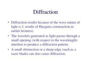
This seminar presentation explores the history and significance of diffraction in crystallography, tracing its roots from early pioneers like W.C. Roentgen and Max von Laue to contemporary applications. It covers the principles behind X-ray, electron, and neutron diffraction methods, their uses in phase identification, and their role in elucidating molecular structures such as myoglobin. The advantages and limitations of each method are discussed, highlighting the critical importance of diffraction techniques in drug development and structural biology today.

E N D
Roles of Diffraction Helen Nguyen Chem. 12B Seminar Presentation Spring ‘05
OUTLINE 1. History of crystallography _ Definition and Greek roots _ 1895, W.C.Roentgen _ 1912, Max von Laue 2. How does it work and what does it use for? _ Useful in phase identification _ Cover the enumeration of the symmetry patterns 3. Methods and diffraction patterns. _ X-ray diffraction _ Electron diffraction _ Neutron diffraction 4. Determination of the first protein using crystallography _ Myoglobin _The experiment _ Brag’s law 5. Advantage and disadvantage 6. Elucidation relative stereochemistry.
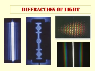
What is Crystallography? • Method uses to study crystals. • Crystallon-solid • Graphein means write • X-ray Diffraction • Electron Diffraction • Neutron Diffraction http://en.wikipedia.org/wiki/Sound http://en.wikipedia.org/wiki/Atomic_nucleus
History • 1895- W.C.Roentgen • 1912- William Lawrence Bragg • Determine the crystalline spacing for a number of substances. • Collimated x-rays to diffract off of different crystal planes. http://www.aip.org/history/esva/catalog/esva/Bragg_Lawrence.html Roentgen http://www.roentgenlauf.de/page5.php?lang=1
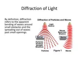
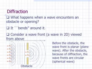
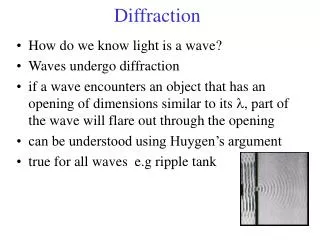
What is Electron Diffraction? • By firing a beam of electrons at a sample and observe their deflection. • The electrons are deflected as waves, as in classical diffraction. Restriction? • Performing of crystal? • Scattered Electrons by…? • Limitation of penetration? • Transmission Electron Microscope (TEM). http://en.wikipedia.org/wiki/Lattice
Neutron Diffraction • Determine the atomic structure of a material. • Found in the atomic nucleus. • All quantum particles can exhibit wave phenomenaassociate with light or sound • Requirements • Interaction? Do not? • Reveal? • Neutrons v.s X-ray http://en.wikipedia.org/wiki/Subatomic_particle • Helium atom (schematic)Showing two protons (red), two neutrons (green) and two electrons (yellow).
X-ray diffraction • Visualize the electron clouds around atoms. • Most important. Pattern? • Analyzing • The lattice is revealed • Bragg's law • Interact with the incoming X-ray photons to diffract them • Myoglobin, Max Perutz and Sir John Cowdery Kendrew http://en.wikipedia.org/wiki/Diffraction http://en.wikipedia.org/wiki/Myoglobin
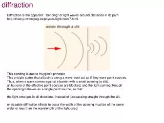
How Brag’s law is used • X-rays hit an atom • Make the electronic cloud move • Rayleigh scattering/Re-radiate waves • Constructive/destructive http://en.wikipedia.org/wiki/Bragg%27s_law
An Experiment Using X-ray Diffraction • Study: a. how oxygen diffused within the protein b. how oxygen is finally released into the surrounding solvent • Substitution of carbon dioxide • Advantage?/Trigger ligand released • The mutant L29F of myoglobin • Conformational changes/ first few hundred picoseconds
Figure 1. 3D (left and center) and 2D (right) representations of the terpenoid, atisane. In the 3D model on the left, carbon atoms are represented by gray spheres, white spheres represent the hydrogen atoms and the cylinders represent the bonds. The model is enveloped in a "mesh" representation of the molecular surface, colored by areas of positive (red) and negative (blue) electric charge. In the 3D model (center), the light-blue spheres represent carbon atoms, the white spheres are hydrogen atoms, and the cylinders in between the atoms correspond to single-bonds. http://en.wikipedia.org/wiki/Molecule
How does X-ray Diffraction Apply to Current Days? • Used to determine how drugs, such as anti-cancer medications • Molecule must be crystallized/why? • Determine a structure/Paintaking? • Diffractometer, a machine that emits a beam of x-rays An X-ray picture (radiograph) taken by Röntgen http://en.wikipedia.org/wiki/X-ray
X-ray Application Cont’ • Diffraction images • What important?/Problem solving--- combination and computational methods • A model made up of atoms is built and refined against the observed data http://en.wikipedia.org/wiki/X-ray X-rays can reveal the details of bones and teeth
Conclusion • X-ray, Neutron, and Electron are the three diffraction methods • X-ray is the method that determined the first protein molecule which is myoglobin by Max Perutz and Sir John Cowdery Kendrew with the use of Brag’s law. • Today X-ray is used to determine how drugs, such as anti-cancer medications
Bibliography • http://en.wikipedia.org/wiki/ • http://web.chem.ucla.edu/~harding/cfqpp/xray30.pdf • http://www.answers.com/topic/x-ray-crystallography • http://www.esrf.fr/UsersAndScience/Publications/Highlights/2003/Materials/MAT15/ • http://en.wikipedia.org/wiki/Atomic_nucleus • http://en.wikipedia.org/wiki/Sound • http://www.roentgenlauf.de/page5.php?lang=1 • http://www.aip.org/history/esva/catalog/esva/Bragg_Lawrence.html • http://en.wikipedia.org/wiki/Lattice • http://en.wikipedia.org/wiki/Subatomic_particle • http://en.wikipedia.org/wiki/Diffraction • http://en.wikipedia.org/wiki/Myoglobin • http://en.wikipedia.org/wiki/Bragg%27s_law • http://en.wikipedia.org/wiki/Molecule http://en.wikipedia.org/wiki/X-ray