Download
1 / 73
860 likes | 1.84k Views
Seronegative Spondyloarthropathies. Goals of the Lecture. Introduce the spondyloarthropathies Recognize AS as the prototypic disease Recognize common clinical and radiologic features and specific features including: Epidemiology Diagnosis Treatment. Seronegative Spondyloarthropathies.

E N D
Goals of the Lecture • Introduce the spondyloarthropathies • Recognize AS as the prototypic disease • Recognize common clinical and radiologic features and specific features including: • Epidemiology • Diagnosis • Treatment
Seronegative spondyloarthropathies (SNSA): A family of diseases • Ankylosing Spondylitis • Reiter’s syndrome/ Reactive arthritis • IBD arthropathy • Psoriatic arthropathy (SNSA variant) • Undifferentiated spondyloarthropathy • Juvenile onset SNSA
SNSA: Group characteristics • Propensity to affect spine, peripheral joints, and periarticular structures • Characteristic extraarticular features • Absence of RF and ANA • Association with HLA B27
SNSA: Group pathology • Sacroiliitis • Osteopenia • Erosions • Peripheral arthritis • Synovial hyperplasia • Pannus • Lymphoid infiltration • Enthesitis • Inflammation at tendinous insertions
Seronegatives AS Reiter’s Psoriatic arthritis IBD SAPHO Acne-associated Intestinal bypass Infections Pyogenic infections Tuberculosis Brucellosis Whipple’s Others Paraplegia Sarcoidosis Hyperparathyroidism Causes of sacroiliitis
Ankylosing spondylitis: Prototype SNSA • Systemic inflammatory • Sacroiliitis is hallmark • X-ray evidence needed for original and modified NY criteria • Clinical spectrum wider than symptomatic sacroiliitis • Atypical
AS: Diagnosis • Diagnostic Criteria • Highly sensitive at early stage of disease • Classification Criteria • Deals with groups of patients • NOT individual patients • Primarily for epidemiologic purposes
Grading sacroiliitis • Grading of radiographs Normal 0 Suspicious 1 Minimal sacroiliitis 2 Moderate sacroiliitis 3 Ankylosis 4
Ankylosing spondylitis(Modified New York classification criteria) 1.LBP at rest for >3 months • improved with exercise • not relieved by rest 2. Limitation of lumbar spine 3. Decreased chest expansion 4. Bilateral sacroiliitis grade 2-4 5. Unilateral sacroiliitis grade 3-4 Definite AS if criterion 4 and any other criteria is fulfilled
Ankylosing spondylitis: Clinical features • Onset in late adolescence/ early adulthood • After age 45 is uncommon • Much more common in men • M:F 3:1 • Clinical/xray features evolve more slowly in women • Skeletal vs. extraskeletal features
AS :Skeletal features • Axial (back pain) • sacroiliitis • spondylitis • Hips/shoulders • Enthesitis • Osteoporosis • Spinal fractures
Ankylosing spondylitisvs. mechanical LBP • Inflammatory/ spondyliticback pain 1. Onset prior to age 40 2. Insidious onset 3. Persistence at least 3 months 4. Morning stiffness 5. Improvement with exercise Need 4/5 criteria
Inflammatory questions • Sensitivity 95-100% • False + 10-15% • mechanical back pain and healthy athletes • low prevalence of AS in population (1-2%) • Positive predictive value is low • 10% false positive
AS: Peripheralskeletal features • Hip and shoulder involvement • May be first symptom • Up to 1/3 patients • More common in juvenile (<16) onset • Flexion contractures at hips
AS: Peripheral skeletal features • Other peripheral joints • Infrequent • Often asymmetric • Transient • Rarely erosive • Resolves without residual deformity
AS: Enthesitis • Enthesitis • Extra-articular or juxta-articular bony pain • Costosternal junctions • Spinous processes • Iliac crests • Greater trochanters • Ischial tuberosities • Tibial tubercles • Achilles tendon insertions • Plantar fascia insertion • Pes anserinus • Epicondylus humeri lateralis
Extraskeletal manifestations • A ortic insufficiency and other cardiac pathology • N eurologic (atlantoaxial subluxation, Cauda equina) • K idney (secondary amyloidosis, chronic prostatitis) • S pine (cervical fracture, spinal stenosis) • P ulmonary (apical lobe fibrosis, restrictive disease) • O cular (anterior uveitis) • N ephropathy (IgA) • D iscitis
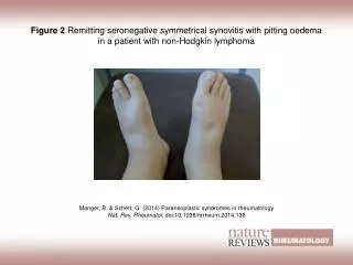
AS: Extraskeletal manifestations • Eye- acute anterior uveitis (25-30%) • Heart- ascending aortitis, AR (3-10%), conduction abnormalities (3%) • Pulmonary- apical fibrosis (rare) • Neurologic- fracture/dislocation. subluxations, cauda equina syndrome
AS: Iritis • Acute anterior uveitis/iritis/ iridocyclitis • Most common ES • 25-30% • Unilateral • Recurrent • Symptoms • Pain • Lacrimation • Photophobia • Blurry vision
AS: Physical examination • Limited range of motion (especially hyperextension, lateral flexion, or rotation) • Spasm/soreness of paraspinal muscles • Positive Schober’s test • Loss of lumbar lordosis • Sacroiliac discomfort
Wiki • The Dimples of Venus (also known as booty dimples, back dimples, or butt dimples) are sagittallysymmetrical indentations sometimes visible on the human lower back, just superior to the gluteal cleft. They are directly superficial to the two sacroiliac joints, the sites where the sacrum attaches to the ilium of the pelvis. • The term "Dimples of Venus", while informal, is an historically accepted name within the medical profession for the superficial topography of the sacroiliac joints. The Latin name is fossae lumbales laterales ('lateral lumbar indentations').These indentations are created by a short ligament stretching between the posterior superior iliac spine and the skin. • Booty dimples are rapidly gaining cultural momentum as a feature men find attractive in women and other men.
Wiki • The dimples of Venus (also known as back dimples) are sagittallysymmetrical indentations sometimes visible on the human lower back, just superior to the gluteal cleft. They are directly superficial to the two sacroiliac joints, the sites where the sacrum attaches to the ilium of the pelvis. • The term "dimples of Venus", while informal, is a historically accepted name within the medical profession for the superficial topography of the sacroiliac joints. The Latin name is fossae lumbales laterales ("lateral lumbar indentations"). These indentations are created by a short ligament stretching between the posterior superior iliac spine and the skin. They are thought to be genetic. • There are other deep-to-superficial skin ligaments, such as "Cooper's ligaments", which are present in the breast and are found between the pectoralis major fascia and the skin. • There is another use for the term "Dimple of Venus" in surgical anatomy. These are two symmetrical indentations on the posterior aspect of sacrum which contain a venous channel too. They are used as a landmark for finding the superior articular facets of the sacrum as a guide to place sacral pedicle screws in spine surgery[1].
1="Vertebra prominens"Spinous process of C72= 2nd Lumbar vertebra3= L4-5 inter vertebral space4= Iliac crests5= Dimples of Venus / Sacroiliac joints / Booty Dimples
AS: Laboratory findings • Elevated ESR (75%) • Elevated CRP • ANA and RF negative • NC/NC anemia (15%) • HLA B27 • No diagnostic or pathognomic tests!
HLA B27 and AS in Caucasian populations • HLA B27 in Americans 8-14% • HLA B27 in African Americans 3% • HLA B27 in AS patients >90% • Prevalence of AS in population 1% • Prevalence of AS in HLA B27+ individuals 2% • Prevalence of AS in B27+ relatives 20% • Prevalence of AS in B27- relatives 0%
AS: Radiologic features • Sacroiliac • Bilateral, symmetric involvement (i.e. erosions, sclerosis, pseudowidening, ossification) • Spine • “Shiny corners”, squaring of the vertebra, ossification of the annulus fibrosus, ankylosis • Hip • Symmetric concentric joint narrowing
AS: Radiographic findings • SI joint- symmetric • Pronounced on iliac side • Erosions/sclerosis • ‘Postage stamp’ serrations • Pseudowidening
More sensitive than XRAY • MRI • CT
Late sacroiliac changes • Calcification, interosseous bridging, and ossification • Bony ankylosis • Osteoporosis
ASRadiographic findings • Vertebral Column • Squaring of vertebrae
Skeletal manifestations • Syndesmophytes • Ossification of the outer layers of the annulus fibrosis • Sharpey’s fibers • Vertical
Late axial disease B A M B O O
AS: Radiographic findings • Enthesitis • Bony erosions • Osteitis (whiskering) of insertions • Ischial tuberosities • Iliac crest • Calcani • Femoral trochanters • Spinous processes
AS: Treatment • Main objectives • Patient education • Early diagnosis • Control pain and suppress inflammation • Daily exercises • Surgical measures (i.e. hip arthroplasty) • Vocational support
AS:Treatment • NSAIDs- pain and stiffness • Sulfasalazine/MTX- peripheral arthritis • Anti-TNF agents- axial and peripheral disease • Oral corticosteroids- little role • Local corticosteroids- recalcitrant enthesopathy
Etanercept in AS (% ASAS Response Week 12)Davis J, et al, Arthritis Rheumatism 2003
Infliximab in AS(% ASAS Response at 24 weeks)van der Heijde D, et al, Arthritis Rheumatism 2005
AS: Summary Age at onset Young adults Sex ratio 3:1 (males to females) Axial disease Virtually 100% Sacroiliitis Symmetric Peripheral joint 25% Eye involvement 25% Infectious triggers Unknown