Concordance of the CKIT Gene with del(9q) in Acute Myeloid Leukemia Research
Investigating the correlation between the CKIT gene and del(9q) in Acute Myeloid Leukemia, exploring mutations in AML samples. Further studies are needed for a comprehensive understanding of genetic factors impacting AML. Differences in sample type and size between studies necessitate more research for precise gene characterization.

Concordance of the CKIT Gene with del(9q) in Acute Myeloid Leukemia Research
E N D
Presentation Transcript
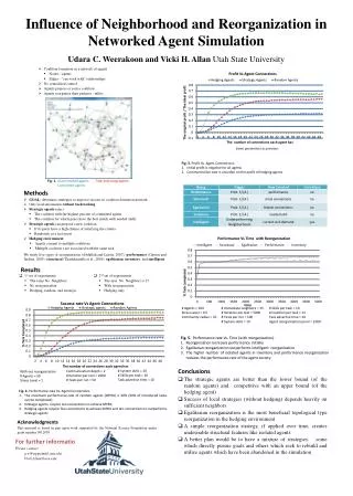
Concordance of the CKIT gene with del(9q) as a cause of Acute Myeloid Leukemia Kyle MarshAuthentic Science Research Program Manchester-Essex Regional High School, Manchester-by-the-Sea, MA 01944 Abstract Concordance of the CKIT gene with del(9q) as a cause of Acute Myeloid Leukemia Kyle MarshManchester-Essex High School, Manchester-by-the-Sea, MA Teacher, Dr. Maria Burgess, Manchester-Essex High School Mentor, Dr. David Sweetser, Mass General Hosp, Boston, MA Acute Myeloid Leukemia (AML) is caused by a combination of various classes of genetic mutations, including tumor suppressor genes, transcription factors/core-binding factors (CBFs), and chromosomal translocations and deletions. One specific deletion on chromosome 9, del(9q), and the translocation t(8;21), have been found together in AML, and it is likely these two mutations cooperate to cause leukemia. Not all cases of del(9q) AML have t(8;21) and it appears that other mutations can cooperate with del(9q). To help better understand how different mutations cooperate to cause leukemia, AML samples with del(9q) were screened for mutations in exons 8 and 17 of the CKIT gene, a cytokine receptor. Of 41 samples, only one mutation was found in exon 8. The low incidence of mutations suggests further studies are necessary. Literature cited Aiello D., Williams J, Majgier-Baranowka H, Patel I, Peet N, Huang J, Lory S, Bowlin L, Moir D. Discovery and Characterization of Inhibitors of Pseudomonas aeruginosa Type III Secretion. Antimicrob. Agents Chemother. 2010. Dayyani F, Wang J, Yeh JR, Ahn EY, Tobey E, Zhang DE, Bernsetien ID, Peterson RT, Sweetser DA. Loss of TLE1 and TLE4 from the del(9q) commonly deleted region in AML cooperates with AML1-ETO to affecxt myeloid cell proliferation and survival. Blood. 2008. Mormann S, Dabisch M, Becker B. Effects of technological processes on the tenacity and inactivation of norovirus genogroup II in experimentally contaminated foods. App Environ. Microbio. 2009.Müller AM, Duque J, Shizuru JA, Lübbert M. Complementing mutations in core binding factor leukemias: from mouse models to clinical applications. Oncogene. 2008.Pollard JA, Alonzo TA, Gerbing RB, Ho PA, Zeng R, Ravindranath Y, Dahl G, Lacayo NJ, Becton D, Chang M, Weinstein HJ, Hirsch B, Raimondi SC, Heerema NA, Woods WG, Lange BJ, Hurwitz C, Arceci RJ, Radich JP, Bernstein ID, Heinrich MC, Meshinchi S. Prevalence and Prognostic Significance of c-KIT mutations in Pediatric Core Binding Factor AML Patients Enrolled on Serial Pediatric Cooperative Trials for De Novo AML. Blood. 2010. Speck and Gilliland. Core-Binding Facts in Haematopoiesis and Leukameia. Nature Reviews Cancer. 2002. Sweetser DA, Peniket AJ, Haaland C, Blomberg AA, Zhang Y, Zaidi ST, Dayyani F, Zhao Z, Heerema NA, Boultwood J, Dewald GW, Paietta E, Slovak ML, Willman CL, Wainscoat JS, Bernstein ID, Daly SB. Delineation of the Minimal Commonly Deleted Segment and Identification of Candidate Tumor-Suppressor Genes in del(9q) Acute Myeloid Leukemia. Genes Chromosomes Cancer. 2005. Verdeguer A. Genetic alterations in children and adolescents with acute myeloid leukaemia. Clin Transl Oncol. 2010. • Conclusions • No mutations found in 41 in exon 17 • 1 mutation in exon 8 • Low incidence of these mutations suggest that further studies are necessary to evaluate the importance of CKIT with concordance to del(9q). • Differences between studies supports further studies to characterize AML samples with the del(9q) for the CKIT gene in order to study its exact relevance in the etiology of AML. • Major difference in our work and Pollard’s was our samples. Pollard solely used pediatric samples, while we used both pediatric and adult AML samples. Pollard also had ~5x more samples than our lab (203 compared to 41), thus, the sample size and type may have affected our results. • Pollard et al. predicted higher incidences of mutations in both exons • AML is a disease controlled by a myriad of both environmental and genetic factors. In AML, del(9q) is found 30% of the time. High incidence of this mutation suggests its potential role in leukemogenesis, Thus, the characterization of each gene associated with del(9q), and of other genes is warranted. Figure 5. Shown is the process of gel electrophoresis. On the left Is a picture of a gel mid-process. DNA’s natural negative charge forces the DNA to run down the gel (through electric currents) towards the positive end of the gel. On the right is a picture of a completed gel – all samples would be strong enough for sequencing except for sample 6, where troubleshooting would be necessary. Figure 1. The CKIT gene. CKIT is a cytokine receptor which, when mutated, has been thought to be involved in the etiology of acute myeloid leukemia. • DNA isolated via column purification and centrifugation • Columns have pores that allow excess material, such as dNTP and Taq, to slide through the membrane and be discarded • DNA is eluded, and exon sent for sequencing • After sequencing, analysis for mutations in exon was performed • MGH “DNA Core” computer software was used • Sequences were obtained through the DNA Core website represented via chromatography peaks each peak representing a different base pair 2 colored bands on top of each other for one nucleotide means there was a possible mutation • Samples were then aligned to check if this was indeed a mutation on a word document, which allowed us to compare nucleotides of each sample. • Materials and Methods • To find mutations in exon 8 and 17, samples were prepared for sequencing. • PCR was used to amplify the section of DNA (exon 8 or 17) of the CKIT gene. • The PCR reaction “master mix” consisted of Taq (DNA polymerase), Taq buffer, primers (to flag the exon), dNTP (nucleotides), and the AML sample. • The reaction was run at 58oC for 35 cycles. • Results • In exon 8, we were looking for a deletion in codons 416-420 • In exon 17, we were looking for point mutations. • We found one mutation for both exons. • Analysis showed this mutation in sample 267, exon 8 only. • Found deletion in codon 419, as previously suggested • Mutations in 2% of the exon 8 samples, compared to 53%, and 0% of the samples had mutations in exon 17, compared to previous study’s 41%. • No significant mutations noted. • Significant polymorphisms in ~5% of the samples at exon 17 • Polymorphism is a synonymous base pair change that is not expressed. • Would be significant in the exon, but insignificant in the intronic sequence, where they were found Acknowledgments I thank Dr. David Sweeter for providing such an excellent opportunity in his laboratory. He was a wonderful mentor who gave me the perfect balance of independence and assistance. He was also a wonderful teacher of the genetics behind AML – I learned a tremendous amount in the laboratory. I would also like to thank the post-docs in Sweetser’s lab who were supportive and available if I had any questions. Supported by the Spaulding Education Fund. Figure 8. Pictured is the del(9q) mutation from three different karyotypes. Figure 6. Once our samples were sequenced, we would access the “DNA Core” website to obtain our samples. Pictured is the exact sequence of one particular sample. Each nucleotide is signified by a different peak. When there was a mutation, there would be a two-toned peak, and a pink “N” where the base pair should be. Figure 2. Pictured is the “master mix” made for PCR. This mix included primers for both exons 8 and 17, Taq (DNA polymerase), Taq buffer, and dNTP (nucleotides). DNA was then added to the mix and run at 58 degrees for 35 cycles. Figure 3. Pictured is the BioRad Polymerase Chain Reaction machine that was utilized for all of our samples. After the reaction was complete, we then ran the amplified samples on a gel in order to see if the DNA sample was readable and ready for sequencing. For further information Please contact km7531@aol.com. More information on this and related projects can be obtained at MGH’s research website, http://www.massgeneral.org/research/researchlab.aspx?id=1196. • DNA samples were then size fractioned via electrophoresis using 1.5% agarose gel. • Ethidum bromide allowed visualization of the DNA. • ~4-5 ml of DNA were inserted into each well. • Pictures of each gel determined if troubleshooting was necessary. • If the DNA band was clear, the sample was sent for sequencing. Figure 7. Shown in exon 8 of the CKIT gene. In sample 267, we found one deletion of codon 419, one of the codons highlighted in the green region that Pollard suggested.