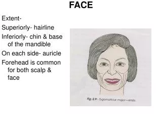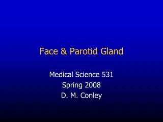
Face & Parotid Gland
420 likes | 1.54k Views
Face & Parotid Gland. Medical Science 531 Spring 2008 D. M. Conley. Face: Regions and Landmarks. Nasolabial sulcus . Skeletal framework = The facial skeleton: Mandible Maxillae Zygomatic bones Nasal bones Lacrimal bones Inferior nasal conchae Vomer.

Face & Parotid Gland
E N D
Presentation Transcript
Face & Parotid Gland Medical Science 531 Spring 2008 D. M. Conley
Skeletal framework = The facial skeleton: Mandible Maxillae Zygomatic bones Nasal bones Lacrimal bones Inferior nasal conchae Vomer Note also the frontal bone contributes greatly to skeletal framework of the face.
Fascia of the Face Superficial fascia is copious and loose – however, there is no discrete layer of deep fascia of the face except …
Deep fascia does exist in the regions of the parotid glands and the masseter muscles. It forms capsules around these structures. The other regions of the face have much subcutaneous tissue, but no deep fascia.
Aging of facial skin Loss of elastic fibers in skin cause permanent wrinkles (e.g., “Crow’s feet” and “worry lines”.
Dynamic duo: Botox & collagen Before After
We will study these muscles in lab, for now think of the facial muscles in groups: Muscles of facial expression: Associated with (1) the forehead, (2) orbit, (3) mouth, and (4) nose.
Buccal fat pad Before After Buccal pad reduction
Blood supply to the face Facial artery Transverse facial a. (from the superficial temporal a.)
Venous drainage of the face Facial vein = A tributary of the internal jugular vein
Lymphatic drainage of the face/scalp • Vessels carrying lymph from the face pass through nodes arranged like a “collar” around the base of the head. • Occipital • Retro-auricular (Mastoid) • Parotid • Submandibular • Submental
Parotid gland: Surface anatomy The parotid has a triangular profile when viewed externally AND in cross-section! Kewl!
Parotid Gland: Relationships Note: (1) External structures bordering the parotid (2) Structures coursing WITHIN the parotid.
Structures coursing within the parotid gland • Facial nerve • Retromandibular vein • External carotid artery • Auriculotemporal nerve (from V3) Superficial Deep
Bell’s Palsy Paralysis of muscles of facial expression due to damage/inflammation of the facial nerve.
Superficial temporal vein Maxillary vein Retromandibular vein Posterior division Anterior division Joins facial v. to form common facial v. A tributary of the internal jugular v. Joins posterior auricular v. to form external jugular v.
External carotid artery Gives off its terminal branches within the parotid Maxillary a. Superficial temporal a.
Auriculotemporal nerve (from V3) Supplies scalp & external ear Carries postganglionic PS fibers that are secretomotor to the parotid.
Parotid (Stensen’s) Duct Penetrates buccal fat pad and buccinator open into the oral cavity opposite the 2nd maxillary molar tooth Note: Its surface anatomy – coursing parallel to the tip of the ear lobule.
Development of the Face [Continued .. From pharyngeal apparatus lecture]
5 primordial structures are crucial to facial development Frontonasal prominence (1) Maxillary prominences (2) Mandibular prominences (2) Primitive mouth (stomodeum)
5 Facial Primordia Frontonasal prominence Maxillary prominences Mandibular prominences From 1st pharyngeal arch
Observe: • The 2 medial nasal prominences fuse = produce the bridge of the nose and the intermaxillary segment. The 2 mandibular prominences fuse = complete the lower jaw. • The 2 lateral nasal prominences fuse with the 2 maxillary prominences (but not with each other)= form the sides (alae) of the nose. The maxillary prominences do not fuse with themselves – they form upper jaw and cheeks.
The lower portion of the fused medial nasal prominences Intermaxillary segment • Gives rise to the: • Philtrum of the upper lip. • Upper incisor teeth & their gums. • Primary palate (region of hard palate just posterior to the upper incisors).
Facial clefts Failure of the embryonic facial prominences to fuse properly Mesenchyme – ectoderm interaction problems = failure of neural crest to migrate properly?
Cleft lip coupled with clefts of the anterior palate or entire palate.
First Arch Syndromes Treacher-Collins Pierre-Robin Cheekbone and mandible hypoplasia, down-slanting palpebral fissures Severe mandibular hypoplasia & cleft palate