FACE
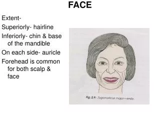
FACE. Extent- Superiorly- hairline Inferiorly- chin & base of the mandible On each side- auricle Forehead is common for both scalp & face. FACE. Skin- Highly vascular Rich in sebaceous gland & sweat gland Laxity- facilitates rapid spread of the oedma

FACE
E N D
Presentation Transcript
FACE Extent- Superiorly- hairline Inferiorly- chin & base of the mandible On each side- auricle Forehead is common for both scalp & face
FACE Skin- • Highly vascular • Rich in sebaceous gland & sweat gland • Laxity- facilitates rapid spread of the oedma • Boils in the nose & ear causes intense pain due to the fixity of the skin to the underlying cartilage • Elastic and thick because the facial muscles are inserted into it.
FACE Superficial fascia- Contents- • Fascial muscles • Vessels & nerves • Fats Deep fascia- absent except over the parotid fascia & over the buccinator where it forms the buccopharyngeal fascia
FACE Fascial muscles- Topographically • Muscles of the scalp- occipitofrontalis • Muscles of the auricle- i) auricularis anterior, ii) auricularis superior & iii) auricularis posterior • Muscles around the eyelids-i) orbicularis oculi, ii) corrugator supercilli & iii) levator palpebrae superiorsis • Muscles of the nose-i) Procerus, ii) Compressor naris, iii) Dilator naris & iv) Depressor septi • Muscles around the mouth- i) orbicularis oris, ii) levator labii superioris alaeque nasi, iii) zygomaticus major, iv)Levator labii superioris, v) Levator anguli oris, vi) zygomaticus minor, vii) Depressor anguli oris viii) Depressor labii inferioris, ix) Mentalis x) Risorius & xi) Buccinator • Muscles of the neck- platysma
FACE Common facial expression- • Smiling & laughing:- zygomaticus major
FACE 2. Sadness:- levator labii superior & levator anguli oris
FACE 3. Grief:- depressor anguli oris 4. Anger:- Dilator naris & depressor septi 5. Frawning:- Corrugator supercilli & procerus 6. Horror, terror & fright:- platysma 7. surprise:- Frontalis 8. Doubt:- Mentalis 9. Grinning:- Risorius 10. Contempt:- zygomaticus minor 11. Closing of mouth:- orbicularis oris 12. Whistling:- buccinator & orbiularis oris
FACE Clinical anatomy- • Frontalis- patient must look upwards without moving the head and look for the horizontal wrinkles of the forehead • Corrugator supercilli- frowning & making the vertical wrinkles of the forehead • Orbicularis oculi- tight closure of the eyes • Orbicularis oris- whistling and pursing the mouth • Dilator of mouth- showing the teeth • Buccinator- puffing the mouth & then blowing forcibly as in whistling • Platysma- forcible pulling of the angle of mouth downwards and backwards forming prominent vertical fold of the skin of the neck. Platysma contracts along with the risorius
FACE Motor nerve supply of the face- • Facial nerve (TZBMC) • Temporal- frontalis, auricularis muscles and orbicularis oculi • Zygomatic- orbicularis oculi • Buccal- muscles of the cheek and upper lip • Marginal mandibular-muscles of the lower lip • Cervical- platysma
FACE Clinical anatomy Infranuclear lesion (Belly’s palsy)- • whole face of the same side gets paralysed. • Face becomes asymmetrical and is drawn up to the normal side. • Affected side is motionless. • Wrinkles disappear from the forehead. • Eyes cannot be closed • While smiling the mouth is drawn to the normal side • During mastication food accumulates in between the teeth and the chick • Articulation of labials is impaired Supranuclear lesions- • usually a part of hemiplegia, only the lower part of the opposite side of the face is paralysed. • Upper part of the frontalis & orbicularis oculi escapes due to its bilateral representation in the cerebral cortex.
FACE Arterial supply of the face- • Facial artery • Transverse facial artery • Arteries accompanying the cutaneous nerves
FACE Facial artery- Extent-Branch of ECA given off in the carotid triangle just above the greater cornua of the hyoid bone. In cervical course it passes through the submandibular region and enters the face region. Course- • Enter face by winding around the base of mandible & by piercing the deep cervical fascia, at the anteroinferior angle of the masster muscle. It can be palpated here and is called the anaesthetist’s artery. • First it runs upwards & forwards to a point 1.25cm lateral to the angel of the mouth. Then it ascends by the side of the nose up to the medial angle of the eye, where it terminates by supplying the lacrimal sac; and by anastomosing with the dorsal nasal branch of the ophthalmic artery. • Lies between the superficial and deep muscles of the face.
FACE Branches- • Anterior branch (large)-i) inferior labial to the lower lip, ii)superior labial to the upper lip and anteroinferior part of the nasal septum &iii) lateral nasal to the ala and dorsum of the nose • Posterior branch-small and unnamed
FACE Transverse facial artery- It is a small branch of the superficial temporal a. After emerging from the parotid gland it runs forward over the masseter between the parotid duct and the zygomatic arch, accompanied by the upper buccal branch of the facial nerv. It supplies the parotid gland and its duct, masseter and the overlying skin and ends by anastomosing with the neighbouring arteries
FACE Veins of the face
FACE Lymphatic drainage- • Preauricular lymph nodes (upper territory)- greater part of forehead,lateral halves of the eyelids, conjunctiva,lateral part of cheek and parotid region • Submandibular lymph nodes ( middle territory)- median part of the forehead, the external nose, the upper lip, lateral part of the lower lip, the medial halves of the eyelids, the medial part of the cheek and greater part of the lower jaw • Submental nodes ( lower territory)- central part of the lower lip and chin
FACE Mucus glands • Labial glands • Buccal glands • Molar glands 1& 2 are numerous and lies in the submucosa of the lips and cheeks 3 lies on the buccopharyngeal fascia around the parotid duct Finally all these opens into the vestibule of the mouth
Eyelids or palpebrae • Upper & lower eyelids Space between this two eye lid- palpebral fissure Two lids fused with each other to form medial & lateral angles or canthi of the eye. At the inner canthi- small triangular space, lacus lacrimalis Lacrimal caruncle(elevated portion in the lacus lacrimalis) Lateral to the caruncle-bulbular conjunctiva is pinched up to form a vertical fold called the plica semilunaris
Eyelids or palpebrae Eyelids- attached at margins of the orbital opening. free edges is broad and has rounded outer lip and a sharp inner lip. Outer lip-2 or more rows of eyelashes or cilia except in the boundary of the lacus lacrimalis. At the point where the eyelashes cease- lacarimal papilla on the summit there is lacrimal punctum. Near the inner lip of the free edge-a row of openings of the tarsal glands
Eyelids or palpebrae Structures- Skin- thin, loose & easily distensible by oedema fluid or blood Superficial fascia- absence of fat, contains palperbral part of the orbicularis oculi Palpebral fascia- two lids forms orbital septum, its thickening forms tarsal plates or tarsi in the lids & palperbral ligs at the angles. Upper tarsus- levator palpebrae superioris & tarsal glands or Meibomian glands embedded in the posterior surface of the tarsi- ducts open in a row behind the cilia Conjunctiva- lines the posterior surface of the tarsus Glands- large sebaceous gland or Zeis’s gland, modified sweat glands or Moll’s gland & sebaceous or tarsal glands or Meibomian glands
Eyelids or palpebrae Blood supply- • Superior & inferior palpebral branches of the ophthalamic a • Lateral palpebral branch of the lacrimal a Veins- ophthalamic and facial vein Lymphatic drainage:- medial halves of the lids -submandibular nodes & lateral halves -preauricular nodes Nerve supply:- Upper eyelid- lacrimal,supraorbital, supratrochlear & infratrochlear nerves Lower eyelid- infraorbital and infratrochlear nerves Lymphatic drainage- medial ½ into the submandibular & lateral ½ into preauricular nodes.
Lacrimal apparatus • Lacrimal gland & its ducts • Conjunctival sac • Lacrimal puncta & lacrimal canaliculi • Lacrimal sac • Nasolacrimal duct
Lacrimal apparatus Lacrimal gland- Serous gland located in lacrimal fossa J shaped, intended by the tendons of the levator palpebrae superioris muscle. a)Orbital part- large & deeper b) Palpebral part- small & superficial
Lacrimal apparatus Nerve supply- lacrimal nerve Arterial supply- ophathalamic a Secretomotor fibres- lacrimatory nucleus----nervus intermedius---- geniculate ganglion—greater petrosal nerve—nerve to pteryoid canal– pterygopalatine ganglion– relay– zygomatic nerve—zygomatotemporal– lacrimal nerve– lacrimal gland
Lacrimal apparatus Conjunctival sac- Potential space between the bulbular conjunctiva and the palpaberal conjunctiva Reflected parted into the eyeball are superior and inferior fronices
Lacrimal apparatus Lacrimal puncta & canaliculi- 10cm long- lacrimal canaliculi 2mm vertical & 8mm horizontal parts Opens into the lateral wall of the lacrimal sac
Lacrimal apparatus Lacrimal sac- 12cm long & 5mm wide- situated in the lacrimal groove behind the medial palpebral lig Upper end blind & lower end continues with nasolacrimal duct Inflammation- dacrocystitis
Lacrimal apparatus Nasolacrimal duct- 18mm long Begins at the lower end of the lacrimal sac, runs downwards, backwards and laterally and opens into the inferior meatus of the nose. Valve of Hanser
Clinical anatomy • Palpebral conjunctiva is examined for anaemia & for conjunctivitis; bulbar conjunctiva for jaundice • Conjunctivitis is one of the commonest diseases of the eye • Trachoma- trachoma virus. Which causes the blindness • Stye or hordeolum is a suppurative inflammation of one of the glands of the Zeis. • Chalazion- inflammation of the tarsal gland • Blepharitis – inflammation of the eyelids specially of the lid margins