Download
1 / 43
640 likes | 1.61k Views
Dr. Ágoston Szél. Surface Anatomy of the Abdomen , Peritoneal Relations. Department of Anatomy , Histology and Embryology Semmelweis University March 5, 2019. Abdominal wall. External oblique abdominal muscle. Internal oblique abdominal muscle. T ransv erse abdominal muscle.

E N D
Dr. Ágoston Szél SurfaceAnatomy of theAbdomen, Peritoneal Relations Department of Anatomy, Histology and Embryology Semmelweis University March 5, 2019
The broadabdominalmuscles ext.obl. abd. m. int. obl. abd. m. iliac crest inguinal lig. lumbar fascia (v) inguinal lig. transv.abd. m. lumbar fascia (v) pubic tubercle inguinal lig.
Rectusabdominalmuscle Pyramidalismuscle
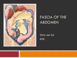
Structure of theabdominalwall superficial fascia skin pectoralis major m. rectusabd. m. aponeurosis of ext. obl. abd. intercostal muscles xyphoid process rectusabd. m. ext. obl. abd. m. linea alba int. obl. abd. m. transv.abd.m. extraperitoneal fat transversalis fascia peritoneum ext. obl. abd. m. obl. abd. m. int. transv.abd. m. transversalis fascia
Rectussheath rectus abdominis muscle inferior epigastric a&v. lateral abdominal muscles urinary bladder vagina external iliac artery. rectum
Arrangement of thefatty and membranouslayers of thesuperficialfascia of theabdominalwall aponeurosis of external oblique muscle superficial fascia membranous layer (Scarpa’s fascia) fatty layer (Camper’s fascia) line of fusion position of penis fascia lata spermatic cord position of scrotum
Arrangement of fasciae in thelowerabdominalwall perineal membrane *fatty layer (Camper’s fascia) symphysis *membranous layer (Scarpa’s fascia) urethra **Colles’s fascia external spermatic, cremasteric, internal spermatic fasciae dartos muscle *superficial fascia **membranous layer of the superficial fascia
Insertion of abdominalmuscles in theloopsystem of the body musculature Rhomboideus Serratus ant. Pectoralis major Int. obl. abd. Gluteus maximus Ext. obl. Abd. Adductors Sartorius
Functions of theabdominalmuscles Movements of the trunk Bending of the trunk (forwards, sideways), rotation of the trunk – in collaboration with the muscles of the back. Stiffening the abdominal wall Fixation of internal organs in position – according to the variation of the content of the stomach, urinary bladder and bowels. Breathing Diaphragm + abdominal muscles. Maintaining the abdominal pressure Expiration, urination, defecation, coughing, sneezing, vomiting, exertion of high muscular force, birth.
Visceraltopography Clavicle Apex of right lung Left lung Left lobe of liver Diaphragm Spleen Right lobe of liver Pancreas, body Stomach Gall bladder Transverse colon Pylorus Duodenum, desc. Ileum Ascending colon Jejunum Cecum Descending colon Ant. inf. iliac spine Urinary bladder
Topographicalcoordinatesystemforsurfaceanatomy (from in front) Medioinguinal plane Medioclavicular (pararectal) plane Jugular notch 1/2 Transpyloric plane (L1) Subcostal plane (L2-3) Supracristal plane (L4) Intercristal distance (L5) Interspinal line 1/2 Symphysis
Topographicalcoordinatesystemforsurfaceanatomy (frombehind) Medioinguinal plane Vertebra prominens Transpyloric plane (L1) Subcostal plane (L2-3) Supracristal plane (L4) Intercristal distance (L5) Interspinal line
Abdominalregionsforphysicalexaminantion Medioclavicular (pararectal) plane Hypochondriacregion EPIGASTRIUM Epigastricregion Subcostal plane McBurney (1/2-1/3) Lateral abd. region umb. MESOGASTRIUM Umbilical region Lanz (1/3) Interspinal plane Ant. sup. iliac spine Inguinal region HYPOGASTRIUM Pubic region
Abdominalregionsforphysicalexamination Mediclavicular (pararectal) plane Hypochondriac r. Diaphragm Epigastric r. Subcostal Lat. abd. r. Umbilical r. Interspinal Inguinal r. Pubic r.
Abdominalregionsforphysicalexamination Anterior axillary fold – navel line Medioclavicular (pararectal) line Murphy 9th rib Monroe-line Morris (2/3) McBurney (1/3) Interspinal l. Lanz (1/3) Sonnenburg: crossing of interspinal line & lateral edge of rectus
Medioinguinal plane Location of visceralorgans in thecoordinatesystem Transpyloric plane (L1) Subcostal plane (L2-3) Supracristal plane (L4) Intercristal distance (L5)
Variations of thetopography of stomach projected ontotheanteriorabdominalwall xyphoid proc. xyphoid proc. cardia cardia fornix 10th rib 10th rib fornix body pylorus pylorus body ant. sup. iliac spine ant. sup. iliac spine hook-shaped (letter J) stomach ptotic (atonic) stomach
Variations of thetopography of stomach projected ontotheanteriorabdominalwall Tp Sc Scr Dcr hook-shaped (letter J) stomach ptotic (atonic) stomach
Projection of internalorgans Lung Heart Stomach Liver Duodenum Gall bladder Descending colon Ascending colon Cecum
Head-zones: irradiation of visceralpain Diaphragm (C4) Heart (T3-4) Esophagus (T4-5) Stomach (T8) Liver, gall bladder (T8-11) Small intestines (T10) Large intestines T11) Urinary bladder (T11-L1) Testis, kidney (T10-L1)
Abdominal guarding, „défensemusculaire” Tensing (hardening) of the abdominal wall muscles is a reflex mechanism. To protect inflamed organs from the pain of pressure upon them. The tensing is detected when the abdominal wall is pressed. Guarding is a characteristic finding in the physical examination for an abruptly painful abdomen (an acute abdomen). It accompanies the inflammation of the inner abdominal (visceral and parietal peritoneal) surface due to appendicitis or diverticulitis. The tensed muscles of the abdominal wall automatically go into spasm to keep the tender underlying tissues from being disturbed.
Position of appendix spine-navel (Monroe’s) line retrocecal position (e) anteroparietal position (b) McBurney’s point (1/2-1/3), Morris (2/3) lumbar position (d) interspinal line ant. sup. iliac spine Lanz’s point (1/3) inguinal lig. pelvic position (a) Ilioinguinal position (c)
Position of appendix Lumbar position (d) Retrocecal position (e) Anteroparietal position (b) Ilioinguinal position (c) Pelvic position (a)
Rovsing-signal: upon vigorously pressing the abdomen counter- clockwise, the pain felt on the appendix region becomes more intensive.
Branches of abdominal aorta esophageal hiatus (Th11) inf. phrenic artery median arcuate ligament aortic hiatus (Th12-L1) coeliac trunk left crus suprarenal artery sup. mesenteric artery gonadal arteries renal artery psoas major muscle lumbar arteries inf. mesenteric artery bifurcation of aorta (L4) common iliac artery median sacral artery bifurcation (sacroiliac joint) external iliac artery internal iliac artery
The prjection of theabdominal aorta and branches inferior vena cava (Th12) coeliac trunk (L1) superior mesenteric artery (L3) inferior mesenteric artery abdominal aorta common iliac a&v. anterior superior iliac spine external iliac a.&v. internal iliac a.&v.
Projection of colon cecum left colic flexure right colic flexure transverse colon ascending colon descending colon ileum distantia cristarum colon sigmoideum cecum rectum appendix medioinguinal plane medioclavicular plane
Projection of liver and gall bladder Medioinguinal plane 5th rib Left lobe 8th rib Right lobe Transpyloric plane Gall bladder Gall bladder
Projection of duodenum and pancreas Superior part (L1) Tail (L1) Descending part (L1-3) Body (L1-2) Duodenojejunal flexure (L1-2) Head (L2) Inferior part (L3) Ascending part (L3-2)
Position of smallintestine Duodenum: horse-shoe shaped, in the upper right quadrant Jejunum: rather vertical, in the upper left quadrant Ileum: rather horizontal, in the lower right quadrant
Topography of spleen (asseenfromtheleft) left lung costo-diaphrag-matic recess diaphragm liver spleen (rib 8/9th-11th) stomach transverse colon
Topography of thelung, pleura, kidney and spleen medioinguinal plane right: 12th rib: upper pole transpyloric plane (L1) left: 12th rib: 1/3-2/3 subcostal plane (L2-3) supracristal plane (L4) intercristal plane (L5)
Posterior projection of kidneys 11th rib 12th rib left kidney right kidney crista iliaca post. sup. iliac spine
Bibliography • Lippert H: Lehrbuch Anatomie, Urban & Fischer, München, 2000 • Mac Kinnon P, Morris J: Oxford Lehrbuch der klinischen Anatomie, Hans Huber, Bern, 1997 • Moore KL, Dalley AF: Clinically Oriented Anatomy, Lippincott, 1999 • Putz R, Pabst R: Sobotta. Alliter, Budapest, 2004. • Patel R: Applied Peritoneal Anatomy, www.myESRorg. • Renz-Polster H, Krautzig S, Braun J: Innere Medizin, Urban & Fischer, München, 2004 • Snell RS, Clinical Anatomy, Little, Brown & Co, Boston, 1995 • Tirkes T et al: Peritoneal Anatomy. Gastrointestinal Imaging, https://doi.org/10.1148/rg.322115032. • https://www.kenhub.com/en/start/anatomy.